
RSS-Feed abonnieren
DOI: 10.1055/a-1730-4529
Gastric pyogenic granuloma: rare entity, usual therapy


Lobular capillary hemangioma, known as pyogenic granuloma, is a benign vascular tumor that generally appears on the skin or in the oral cavity but rarely occurs in the gastrointestinal tract, where it can cause bleeding [1]. Although gastric pyogenic granuloma is rarely reported in the literature, (up to 2016, approximately 50 cases of gastrointestinal pyogenic granuloma in the English literature had been indexed on MEDLINE, including a few cases of gastric involvement), the actual incidence is probably higher [2] [3].
The endoscopic appearance of pyogenic granuloma is usually a single polypoid lesion, smooth and ulcerated; the color ranges from bluish to reddish with a superficial white or opaque film covering. Resection of pyogenic granuloma is necessary in patients with anemia, but post-resection bleeding is a potential complication. The lesions typically involve the mucosa but may extend to the deep layers; thus, preoperative endoscopic ultrasonography is recommended [1] [4] [5].
Histopathologically, pyogenic granuloma is a hemangioma characterized by a lobule-like growth of capillaries with enlarged vascular endothelial cells and inflammatory cell infiltration in the stroma. Granulation tissue may also be present; the main pathological differential diagnosis of pyogenic granuloma includes bacillary angiomatosis, Kaposi’s sarcoma, or inflammatory and/or hyperplastic polyps [1] [3].
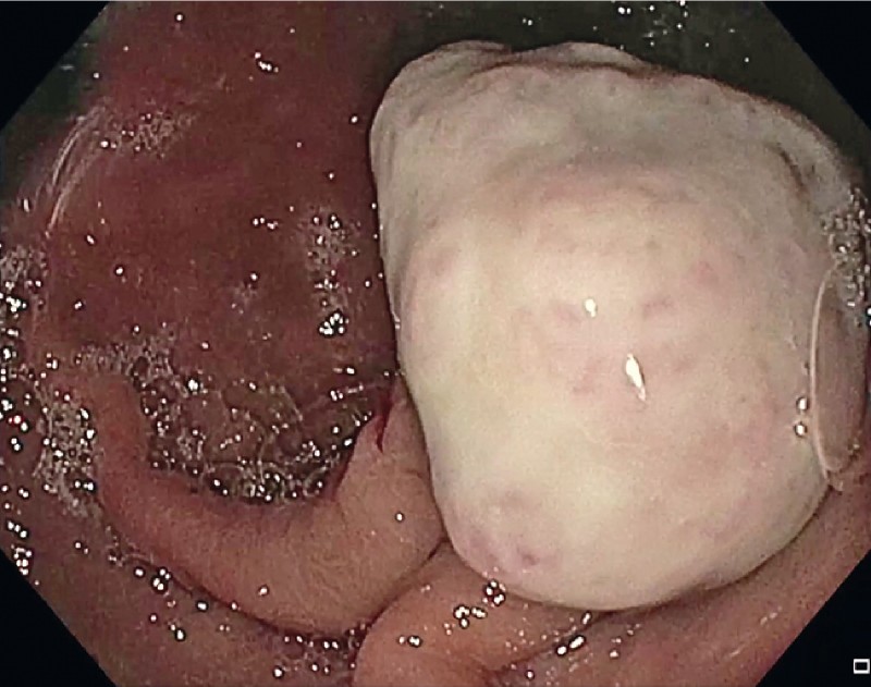
We report a case of a 78 year-old Caucasian woman with a medical history significant for ibuprofen use admitted to our department for anemia and melena requiring transfusion. Esophagogastroduodenoscopy revealed a nearly 20-mm pedunculated polyp, strongly hyperemic with a superficial white film, in the gastric body ([Fig. 1]). Endoscopic ultrasonography showed mucosal involvement without deep infiltration ([Fig. 2]).

We removed the polyp using endoscopic mucosal resection, lifting the lesion with a solution of indigo carmine and epinephrine; in addition, multiple clips were used to close the defect to prevent bleeding ([Video 1]). Histology demonstrated foveolar hyperplasia and lobulated capillary hemangioma, characteristic of pyogenic granuloma ([Fig. 3], [Fig. 4]). Her refractory anemia improved after the procedure.
Video 1 Gastric pyogenic granuloma effectively removed by endoscopic snare resection.
Qualität:


Endoscopy_UCTN_Code_CCL_1AB_2AD_3AC
Endoscopy E-Videos is an open access online section, reporting on interesting cases and new techniques in gastroenterological endoscopy. All papers include a high quality video and all contributions are freely accessible online. Processing charges apply (currently EUR 375), discounts and wavers acc. to HINARI are available.
This section has its own submission website at https://mc.manuscriptcentral.com/e-videos
Publikationsverlauf
Artikel online veröffentlicht:
04. Februar 2022
© 2022. Thieme. All rights reserved.
Georg Thieme Verlag KG
Rüdigerstraße 14, 70469 Stuttgart, Germany
-
References
- 1 Hayashi Y, Hosoe N, Takabayashi K. et al. Clinical and endoscopic characteristics of pyogenic granuloma in the small intestine: a case series with literature review. Intern Med 2020; 59: 501-505
- 2 Katsurahara M, Kitade T, Tano S. et al. Pyogenic granuloma in the small intestine: a rare cause of obscure gastrointestinal bleeding. Endoscopy 2015; 47: E133-E134
- 3 Val-Bernal JF, Mayorga M, Cagigal ML. et al. Gastric pyogenic granuloma: report of two cases and review of the literature. Pathol Res Pract 2016; 212: 68-71
- 4 Reichert MC, Schuster M, Kim YJ. et al. Recurrent gastrointestinal bleeding due to multiple pyogenic granulomas in the stomach. Am J Gastroenterol 2017; 112: 833
- 5 Meeks MW, Kamal UM, Hammami MB. et al. Gastrointestinal pyogenic granuloma (lobular capillary hemangioma): an underrecognized entity causing iron deficiency anemia. Case Rep Gastrointest Med 2016; 2016: 4398401