Keywords
nevus lipomatosus superficialis - sebaceous trichofolliculoma -
Demodex
- hamartoma
Introduction
Nevus lipomatosus superficialis (NLS) is a benign cutaneous hamartoma in which mature
adipose tissue is deposited ectopically between collagen bundles in the dermal layer,
as observed on histopathological examination. It was first reported by Hoffmann and
Zurhelle in 1921.[1] The lesions have two clinical manifestations: (1) The classical type is a zosteriform
pattern consisting of nontender, soft, skin-colored or yellow papules, nodules, or
plaques that are present at birth or appear during the first three decades of life,
and (2) the solitary type is a single sessile, dome-shaped papule or nodule that appears
in the third to sixth decades of life.[2] Malignant transformation has not been reported. Surgical excision is curative, and
no recurrence has been reported.
We describe a case of NLS that manifested with an unusual combination of histopathological
findings of sebaceous trichofolliculoma and Demodex infestation. We then discuss the findings of a literature review and summarize the
clinical manifestations.
Case
The patient, a 41-year-old man, presented with a 35 × 20 mm, cerebriform nodular skin
lesion in the posterior region near the right auricle, and satellite papules on the
posterior surface of the auricle; these asymptomatic lesions had been present since
the age of 14 years. The lesions became prominent after the age of 16 years and gradually
enlarged after the age of 29 years. The patient sought treatment for cosmetic reasons
alone. Only the main lesion, including the subcutaneous tissue, was excised elliptically.
To achieve approximation of both skin margins, the skin flaps were advanced after
the undermining procedure. Preoperative and postoperative findings are illustrated
in [Fig. 1]. By 3 months after surgery, the lesion had not recurred.
 Fig. 1 Preoperative appearance. The lesion (3 × 1.5 cm) was noted in the posterior region
near the right auricle over the auriculotemporal sulcus. (A) An elliptical excision line was designed. (B) After surgery, the skin margins of the excised wound were approximated and well healed
by postoperative day 15.
Fig. 1 Preoperative appearance. The lesion (3 × 1.5 cm) was noted in the posterior region
near the right auricle over the auriculotemporal sulcus. (A) An elliptical excision line was designed. (B) After surgery, the skin margins of the excised wound were approximated and well healed
by postoperative day 15.
Gross and Histopathological Findings
The excised skin specimen (52 × 23 mm) contained a 25 × 25 skin-colored nodule with
a cerebriform surface. Serial sectioning revealed yellowish-white contents, lobulation,
and deposition of adipose tissue in the dermis ([Fig. 2]). Histopathological examination revealed that the nodule was composed of folliculosebaceous
material and that groups of mature adipose tissue occupied the whole dermis ([Fig. 3A, B]). Demodex mites were also present in follicles and sebaceous glands with perifollicular inflammation
([Fig. 3C]). The pathological diagnosis was NLS with sebaceous trichofolliculoma and Demodex infestation with inflammation.
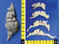 Fig. 2 Gross photographs of serial sections, showing skin-colored pedunculated multiple
nodules with cerebriform surfaces (A) and yellowish-white lobulated nodules and underlying adipose tissue in the dermis
(B).
Fig. 2 Gross photographs of serial sections, showing skin-colored pedunculated multiple
nodules with cerebriform surfaces (A) and yellowish-white lobulated nodules and underlying adipose tissue in the dermis
(B).
 Fig. 3 The nodule was composed of many sebaceous lobules with hair follicles (A) and underlying groups of mature fat cells among the dermal collagen (B). Hematoxylin and eosin ([H&E]; magnifications, 12.5 [A] and 40 [B]). (C)
Demodex mites were found in follicles with perifollicular inflammation and in sebaceous glands
(inset) (H&E; magnifications, 100 [C] and 400 [inset]).
Fig. 3 The nodule was composed of many sebaceous lobules with hair follicles (A) and underlying groups of mature fat cells among the dermal collagen (B). Hematoxylin and eosin ([H&E]; magnifications, 12.5 [A] and 40 [B]). (C)
Demodex mites were found in follicles with perifollicular inflammation and in sebaceous glands
(inset) (H&E; magnifications, 100 [C] and 400 [inset]).
Literature Review
The number of published articles about NLS has increased significantly since 2012
([Fig. 4]). Awareness of NLS has probably increased in many countries. We conducted a literature
review, for which we searched PubMed with the keyword “NLS.” The search was limited
to articles written in English and whose full text was available. We analyzed the
following data: year of report, nation of corresponding author, sex of patient, age
at onset, duration of disease, location of lesion, type of lesion, associated symptoms,
pathological findings, and treatment. Among the countries of the authors ([Fig. 5]), India, the Republic of Korea, and the United States were most common. In total,
112 articles about 149 cases were selected from the PubMed search. Of the 149 patients,
78 were male and 71 were female. We found no sex difference in the prevalence of NLS.
In 114 cases, the lesions were the classical type; in 33 cases, the solitary type.
Two patients had both classical and solitary lesions ([Fig. 6]).
 Fig. 4 A case of nevus lipomatosus superficialis (NLS) was first reported in 1921. We analyzed
112 articles identified in the PubMed search. The number of reports of NLS has increased
gradually since 2003.
Fig. 4 A case of nevus lipomatosus superficialis (NLS) was first reported in 1921. We analyzed
112 articles identified in the PubMed search. The number of reports of NLS has increased
gradually since 2003.
 Fig. 5 The 112 selected articles concerned cases reported from all continents and 26 nations.
We found no geographic preponderance.
Fig. 5 The 112 selected articles concerned cases reported from all continents and 26 nations.
We found no geographic preponderance.
 Fig. 6 Nevus ipomatosus superficialis (NLS) has two manifestations: classical and solitary.
(A) Among the selected articles, the classical type was reported more commonly than the
solitary type. Two patients had both classical and solitary lesions. (B) The lesions were located in five regions: head and neck, trunk, pelvis and genitalia,
upper extremities, and lower extremities. Classical lesions appeared predominantly
on the pelvis and trunk. The solitary type appeared most frequently on the pelvis
and lower extremities.
Fig. 6 Nevus ipomatosus superficialis (NLS) has two manifestations: classical and solitary.
(A) Among the selected articles, the classical type was reported more commonly than the
solitary type. Two patients had both classical and solitary lesions. (B) The lesions were located in five regions: head and neck, trunk, pelvis and genitalia,
upper extremities, and lower extremities. Classical lesions appeared predominantly
on the pelvis and trunk. The solitary type appeared most frequently on the pelvis
and lower extremities.
The lesions were located in five regions: head and neck, trunk, pelvis and genitalia,
upper extremities, and lower extremities. Classical lesions were predominantly on
the pelvis and trunk, and solitary lesions were frequently on the pelvis and lower
extremities ([Fig. 6]).
With regard to onset, findings in previous reports[2] were consistent with ours: the classical type usually was present at birth or appeared
in the first three decades of life, and the solitary type appeared in the third to
fifth decades of life ([Fig. 7]). In 28 patients (18%), classical and solitary lesions were congenital.
 Fig. 7 Onset of lesion. The classical type of nevus lipomatosus superficialis (NLS) usually
was present at birth or appeared in the first three decades of life. The solitary
type appeared in the third to fifth decades of life. In 28 cases (18%), the classical
and solitary types were congenital.
Fig. 7 Onset of lesion. The classical type of nevus lipomatosus superficialis (NLS) usually
was present at birth or appeared in the first three decades of life. The solitary
type appeared in the third to fifth decades of life. In 28 cases (18%), the classical
and solitary types were congenital.
“Duration of the disease” means the time interval between the patient's awareness
of the lesion's presence and the visit to the clinic. Among durations of less than
20 years, five were most common ([Fig. 8]).
 Fig. 8 “Duration of the disease” means the time interval between the patient's awareness
of the lesion's presence and the visit to the clinic. Among durations of less than
20 years, five durations were most common. This suggested that nevus lipomatosus superficialis
was typically present for more than 5 years and that the lesion increased gradually
in size.
Fig. 8 “Duration of the disease” means the time interval between the patient's awareness
of the lesion's presence and the visit to the clinic. Among durations of less than
20 years, five durations were most common. This suggested that nevus lipomatosus superficialis
was typically present for more than 5 years and that the lesion increased gradually
in size.
Discussion
In the United States, the Food and Drug Administration defines a rare disease as any
disease that affects fewer than 200,000 Americans. In Europe, a disease is defined
as rare if it affects fewer than 1 per 2,000 people.[3] Until now, no more than 200 cases of NLS were reported, according to our PubMed
search; thus, NLS could have been classified as a rare disease. Data on prevalence
will be required in the near future.
Three patients with NLS had relatives with the condition.[4]
[5] In one case of NLS, the patient had a 2p24 deletion; the authors suggested that
the chromosomal abnormality played a role in NLS.[6]
NLS has been described as asymptomatic. According to our literature review, however,
10 patients (6.7%) presented with symptoms such as itching, pain, foul odor, and rhinitis.
One patient had dyspnea as a result of an intranasal lesion. According to another
report, compressive neuropathy of the ulnar nerve was caused by firm subcutaneous
nodules of thickened fibrotic dermis containing adipose tissue.[7] Systemic abnormalities and malignant changes have not been associated with NLS.
Several combinations of lesions have been reported. Comedones, abnormal hair growth
or alopecia, and acanthosis most frequently accompanied NLS. Combinations of NLS with
trichofolliculoma, folliculosebaceous hamartoma, or perifollicular fibroma were rare.
In two cases, NLS was accompanied by intramuscular lipoma.[8]
[9] In one case, NLS was accompanied by a polypoid type of basal cell carcinoma of the
scalp.[10] In our patient, NLS and sebaceous trichofolliculoma were present; in our literature
review, we found only one report in which NLS was accompanied by a sebaceous trichofolliculoma.[11]
Sebaceous trichofolliculoma has hair follicles with sebaceous gland components, which
are epithelial components. However, there are two other, similar lesions that should
be differentiated from sebaceous trichofolliculoma histopathologically: trichofolliculoma,
which has only a follicular structure, and folliculosebaceous hamartoma, which contains
not only epithelial components of follicles and sebaceous components but also mesenchymal
structures of vessels, cartilage, and other tissues.
Plastic surgeons are not familiar with NLS. It is often misdiagnosed as a common skin
lesion such as soft fibroma, skin tag (acrochordon), neurofibroma, or lipoma. The
differential diagnosis of the classical type includes mucinous nevus,[12] sebaceous nevus, hemangioma, hairy nevus, lymphangioma, and focal dermal hyperplasia
(Goltz syndrome). The solitary type is often confused with skin tags, solitary neurofibromas,
and solitary lipofibromas.[13]
Treatment is usually not necessary except for cosmetic reasons. Surgical resection
is curative, and no recurrence has been reported. Other treatment modalities have
been attempted: CO2 laser treatment,[14]
[15] cryotherapy,[16] intralesional phosphatidylcholine injection,[17] topical corticosteroid application,[18] and electrodessication.[19] However, because the lesion is large, reconstructing the defect after lesion removal
is difficult. Physicians should be aware of this rare disease because early detection
allows for less invasive resection of the lesion and more conservative reconstruction
of the defect.


