Keywords
hemiclamshell - aortic arch aneurysm - ductus arteriosus
Introduction
The anatomical relations of posterior aortic arch aneurysms force us to address the
issue of the proper surgical approach.[1] In fact, the incision should guarantee a safe dissection of the supra-aortic trunks
and the descending aorta and also allow the cannulation for extracorporeal circulation
(ECC). Protecting the cerebral flow and circulation downstream to the aortic clamping
is yet another challenge that this pathology dictates. Another challenge is to preserve
both the left phrenic and recurrent laryngeal nerves, while sparring a maximum of
the chest wall muscles. We present here a left hemiclamshell approach for posterior
aortic arch aneurysms.
Techniques and Results
We report our strategy regarding two cases of posterior aortic arch aneurysms. Both
cases were discovered incidentally and involved young male patients (36 and 26 years
old).
For patient 1, the involvement of the left subclavian artery made endovascular aneurysm
repair (EVAR) impossible ([Fig. 1]). For patient 2, it was the short landing zone that contraindicated EVAR (landing
zone < 1 cm; [Fig. 2]).
 Fig. 1 Computed tomography scan of patient 1. A,aneurysm ; LSCA,left subclavicular artery.
 Fig. 2 Computed tomography scan of patient 2. A, aneurysm; LSCA, left subclavicular artery.
Hence we proceeded with therapy by direct open surgical repair of those two posterior
aortic arch aneurysms using the left hemiclamshell approach.
The patients were placed in supine position with a log lifting their left hemithorax.
The left lung was excluded from ventilation. The approach consisted of a partial vertical
median sternotomy extended into an anterior thoracotomy in the fourth intercostal
space. The left mammary pedicle was ligated and severed near the fourth intercostal
space. To facilitate exposure, we cut through the posterior arch of the first rib.
The posterior arch of the left first rib is transected from inside the thorax with
a standard rib cutter.
The surgery followed with the dissection of the supra-aortic trunks, the descending
aorta, the left phrenic nerve, and the left recurrent nerve ([Fig. 3]). When working in close proximity with the aneurysm, extracorporeal circulation
was used.
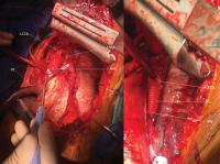 Fig. 3 Patient 2, intraoperative view. AAC, ascending aota cannulation; DG, Dacron graft;
LCCA,left common carotid artery; LSCA, left sub-clavicular artery; PN, phrenic nerve;
VC,venous cannula.
Fig. 3 Patient 2, intraoperative view. AAC, ascending aota cannulation; DG, Dacron graft;
LCCA,left common carotid artery; LSCA, left sub-clavicular artery; PN, phrenic nerve;
VC,venous cannula.
ECC consisted of an atriocaval cannulation through the right atrium and a double arterial
cannulation (femoral artery and descending aorta [[Fig. 3]]). The technique answered to the need for protecting the cerebral flow and the circulation
downstream to the aortic clamping during luxating the heart. No cardioplegia was used
during ECC. Proximal aortic clamping was done between the left carotid artery and
the left subclavian artery. Distal clamping was done on the descending aorta, a few
centimeters downstream from the aneurysm. The left subclavian artery was clamped separately.
For patient 1, the aneurysm sack was spared but two intercostal arteries were ligated,
whereas patient 2 benefited from a total resection of the aneurysm. Since no intercostal
artery emerged from the aneurysm, we did not have to ligate any of them.
Dacron prostheses were used to reestablish aortic continuity (size 26 mm for patient
1 and size 20 mm for patient 2). Reimplantation of the left subclavian artery was
necessary for patient 1 ([Fig. 4]).
 Fig. 4 Patient 1, intra operative view and the rib cutter. LSCAR,left sub-clavicular artery
reimplantation.
Fig. 4 Patient 1, intra operative view and the rib cutter. LSCAR,left sub-clavicular artery
reimplantation.
Cardiopulmonary bypass and aortic clamping times were 142 and 108 minutes for patient
1 and 86 and 49 minutes for patient 2, respectively.
Patient 2 exited the operating room with two analgesic catheters on both sides of
the incision.
Surgical outcomes consisted of a transient diaphragmatic paresis in patient 1. No
laryngeal paralysis was observed. Postoperative computed tomography did not show any
anastomotic pseudoaneurysms. Wounds were clean and no skin infection was noted ([Fig. 5]).
 Fig. 5 Postoperative wound.
Discussion
In both the cases, the suspected etiology is ductus arteriosus aneurysm. There was
no history of thoracic trauma or infection to contradict this hypothesis.[2] Unlike the case by Buechner et al,[3] EVAR was not practicable for the reasons listed above (i.e., the damage of the left
subclavian artery for patient 1 and the short landing zone for patient 2).
Left hemiclamshell approach allows double arterial cannulation (femoral artery and
descending aorta) for the purpose of protecting the cerebral flow and the circulation
downstream the aortic clamping during luxating the heart for the dissection of the
posterior mediastinum. It also offers a satisfying operative view of all the key anatomical
structures to operate in good and secure conditions, while limiting the risks of nerve
lesions (phrenic and recurring nerves) and muscle infringement (latissimus dorsi,
serratus anterior, trapezius, and rhomboid major). Postoperative pain and rehabilitation
ensue from the respect of these structures.
For patient 2, posterolateral thoracotomy could have been contemplated. However, the
previous requirements (nerve and muscle sparring) and the need for perfect access
to the aortic cross between the left carotid artery and the left subclavicular artery
kept us away from that approach.
In conclusion, the left hemiclamshell approach for posterior aortic arch aneurysm
seems to be a good compromise regarding all the requirements listed above.


