
Subscribe to RSS
DOI: 10.1055/s-0041-1730335
Spinal Cord Paracoccidioidomycosis: Case Report
Paracoccidioidomicose na medula espinhal: Relato de casoAbstract
Paracoccidioidomycosis (PCM) is a systemic mycosis caused by fungi Paracoccidioides brasiliensis and Paracoccidiodes Lutzii. Its distribution is limited to subtropical regions of Central and South America, where it is endemic, and Brazil accounts for ∼ 80% of the reported cases. Even in endemic zones, its incidence is low, ranging from 3 to 4 new cases per million to 1 to 3 new cases per 100 thousand inhabitants per year. Granulomas in the spinal cord are rare, and they account for 0,6% of all cases of systemic PCM. The authors report a case of a woman with crural paraparesis caused by dorsal spinal cord PCM granulomasin T7-T8 and T8-T9, with no evidence of systemic disease. The patient was submitted to microsurgery, with total excision of the lesions, and is experiencing positive neurological recovery. Though rare, PCM intramedullary granulomas must be considered in differential diagnosis of the tumoral expansive process of the spinal cord, especially in patients coming from endemic rural zones.
#
Resumo
A paracoccidioidomicose (PCM) é uma micose sistêmica causada pelos fungos Paracoccidioides brasiliensis e Paracoccidioides Lutzii. A doença é endêmica nas regiões subtropicais das Américas do Sul e Central, sendo o Brasil responsável por aproximadamente 80% dos casos relatados. A sua incidência, até mesmo em zonas endêmicas, é baixa, e varia de 3 a 4 casos novos por milhão até 1 a 3 casos novos por 100 mil habitantes ao ano. Os granulomas intramedulares são raros, e acometem 0,6% dos indivíduos com PCM. Os autores relatam o caso de uma paciente de 81 anos com paraparesia crural devido a granulomas intramedulares de PCM em T7-T8 e T8-T9, sem evidências de doença sistêmica. A paciente foi submetida a microcirurgia, com boa evolução pós-operatória. Embora raros, os granulomas intramedulares de PCM devem ser considerados no diagnóstico diferencial das lesões da medula espinhal, especialmente naqueles pacientes provenientes de zonas rurais endêmicas.
#
Introduction
Paracoccidioidomycosis (PCM) is a systemic granulomatous chronic mycosis caused by fungi Paracoccidioides brasiliensis and Paracoccidioides Lutzii.[1] [2] [3] It is endemic in the subtropical areas of South and Central America,[1] [4] [5] and Brazil accounts for ∼ 80% of the reported cases, followed by Colombia, Venezuela, Argentina, and Peru.[2] [3] [5] [6] It occurs more often in rural areas of the Brazilian south, southeast and midwest, especially in the states of São Paulo and Minas Gerais.[1] [2] [7] [8]
After being inhaled, the Paracoccidioides fungi cause pulmonary infection that is often subclinical in immunocompetent individuals. And, should the occasion arise, such fungi might spread to other organs, either through hematological or lymphatic routes, in special to the oropharynx, the mucocutaneous tissue, the lymph nodes, the adrenal glands, the liver, as well as to the central nervous system (CNS).[1] [2] [3] [9]
Involvement of the CNS is rather variable, occurring in 0% to 27% of the series of patients with PCM.[1] [2] [3] [6] The granulomatous form predominates in 96% of the cases, occurring especially in cerebral hemispheres.[1] [10] [11] Intramedullary granulomas are rare, affecting 0,6% of PCM cases and 4% of cases of neuroparacoccidioidomicosys.[1] [9] Reviewing the Medline and LILACS databases, we identified 19 reports of patients with intramedullary granulomas.[6] [7] [9] [12] [13] [14] [15] [16] [17] [18] [19] [20] [21] [22] [23] Due to their rarity, we herein report the case of a patient with two dorsal intramedullary lesions which were approached surgically.
#
Case Report
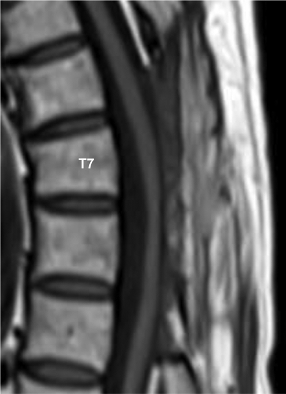
An 81-year-old woman presented with a history of progressive weakening of muscular strength on the lower limbs which had started one month before the consultation. Four months before, she had presented herpes zoster on the dorsal region of the skin at the level of the roots of T4 and T5 to the right. There was no other neurologic symptomatology. The patient had been a smoker since she was an adolescent, and had systemic arterial hypertension. She had lived and worked in a rural area of the state of Rio Grande do Sul, Brazil. Upon neurological examination, the patient presented crural paraparesis with grade force II, bilateral Babinsky, and sensorial level in T9. She was investigated with magnetic resonance imaging (MRI), and two intramedullary spinal cord images were suggested granuloma or metastasis in T7-T8 and T8-T9 ([Fig. 1]). Due an increase in the weakening of muscular strength on the lower limbs, the patient was submitted to laminectomy from T6 to T9, as well as to a total microsurgical excision of the lesions ([Fig. 2]). Intraoperative ultrasonography was performed to locate the intramedullary granulomas ([Fig. 3]). The histopathological examination was compatible with PCM ([Fig. 4]). We used dexamethasone 16 mg/day preoperatively and postoperatively.


The patient was referred to an infectious disease specialist, performing a chest computed tomography, which revealed a lump in the posterior segment of the right upper lobe, and another one in the left upper basal segment. Furthermore, small calcified granulomas were observed, spread bilaterally in the lung parenchyma. There were no signs suggestive of either skin or oral lesions, and no involvement of other organs and systems. The patient was treated with sulfamethoxazole and trimethoprim.
A year after the surgical treatment, the patient presented a positive evolution, and was capable of ambulation with the aid of metal bars.
#
Discussion
Paracoccidioidomycosis is not a disease of compulsory notification; therefore, there is no precise data about its incidence and prevalence in Brazil. It is believed that its incidence in endemic areas may vary from 3 to 4 new cases per million to 1 to 3 new cases per 100 thousand inhabitants per year.[1] [2] [3] [9] However, recent records of incidence in the state of Rondônia, Brazil, report 9,4 cases per 100 thousand inhabitants.[10] Clinical findings generally occur between the ages of 30 to 50 years, old and male patients are more affected in the proportion of 10 to 15 for 1 female.[1] [2] [3] [9]
Once inhaled, the fungus may be either destroyed in the lung parenchyma by phagocytic cells or multiply and produce an infection source, forming a primary complex. Such lesions may either recede in immunocompetent individuals or the fungi contained in these complexes may spread to other organs, causing the acute form of the disease. Many individuals remain with the primary scarring complex containing viable fungi, called quiescent lesions, which may be reactivated and evolve to chronic PCM many years after the initial infection. In the tissues, reactivity of the host induces an inflammatory response and the formation of granulomas. In this respect, granuloma represents an immune response triggered by the host to the wall components released by the offending agent.[1] [2] [3] [6] [9] Involvement of the CNS is always secondary to the primary focus, and there is usually a widespread illness affecting multiple organs. However, simultaneous involvement of other organs or systems may not occur.[1] [6] [9] [15] Smoking and alcoholism are often associated.[2]
One of the reasons for the difficulty in establishing a diagnosis in patients without systemic disease is that the neuroradiological characteristics of the lesions are not specific, being impossible to differentiate them from granulomas caused by tuberculosis, toxoplasmosis, cysticercosis, cryptococcosis or even glial and metastatic neoplasia.[1] [11] [12] [13] [14] [16] [19] [22] [23] The hypothesis of granuloma must be considered when multiple lesions are observed.[13] [23] Besides intramedullary spinal cord involvement, intradural extramedullary granulomas may occur, as well as epidural bone lesions involving the spinous process, the lamina and the vertebral bodies, causing osteomyelitis and spondylodiscitis.[24] [25] [26] [27] [28]
There is evidence that the concomitant use of corticosteroids with antifungal therapy can reduce the inflammatory process in patients with PCM.[2] The drugs most used in the antifungal treatment are itraconazole, sulfamethoxazole/trimethoprim, and amphotericin B.[2] Microsurgical treatment is recommended in those patients who present neurological signs of spinal cord lesions.[1] [2] [6] [9] In the case herein reported, intraoperative ultrasonography facilitated the identification of the lesions and the surgical approach.
Although there was an assumption of granuloma, the PCM diagnosis came to us as a surprise. Even though PCM is a rare disease, it must be considered in the differential diagnosis of intramedullary and intracranial expanding processes, especially in those patients coming from endemic rural areas and, furthermore, awareness of this disease must be raised, to make the diagnosis easier and enable physicians to provide early treatment.
#
#
Conflict of Interests
The authors have no conflict of interests to declare.
-
References
- 1 de Almeida SM. Central nervous system paracoccidioidomycosis: an overview. Braz J Infect Dis 2005; 9 (02) 126-133
- 2 Shikanai-Yasuda MA, Mendes RP, Colombo AL. et al. Brazilian guidelines for the clinical management of paracoccidioidomycosis. Rev Soc Bras Med Trop 2017; 50 (05) 715-740
- 3 Moreira APV. Paracoccidioidomicose: histórico, etiologia, epidemiologia, patogênese, formas clínicas, diagnóstico laboratorial e antígenos. Bol Epidemiol Paul 2008; 5 (51) 11-24
- 4 Fagundes-Pereira WJ, Carvalho GTC, Góes AM. et al. Paracoccidioidomicose do sistema nervoso central. Arq Neuropsiquiatr 2006; 64 (02) 269-276
- 5 Pedroso VSP, Vilela MC, Pedroso ERP, Teixeira AL. Paracoccidioidomicose com comprometimento do sistema nervoso central: revisão da literatura. Rev Bras Neurol 2008; 44 (03) 33-40
- 6 Paniago AMM, de Oliveira PA, Aguiar ESA. et al. Neuroparacoccidioidomycosis: analysis of 13 cases observed in an endemic area in Brazil. Trans R Soc Trop Med Hyg 2007; 101 (04) 414-420
- 7 de Moura LP, Raffin CN, del Negro GM, Ferreira MS. Paracoccidioidomicose evidenciando comprometimento medular tratada com sucesso por fluconazol. Arq Neuropsiquiatr 1994; 52 (01) 82-86
- 8 de Souza SP, Jorge VM, Xavier MO. Paracoccidioidomycosis in southern Rio Grande do Sul: a retrospective study of histopathologically diagnosed cases. Braz J Microbiol 2014; 45 (01) 243-247
- 9 de Almeida SM, Queiroz-Telles F, Teive HAG, Ribeiro CE, Werneck LC. Central nervous system paracoccidioidomycosis: clinical features and laboratorial findings. J Infect 2004; 48 (02) 193-198
- 10 Vieira GdeD, Alves TdaC, Lima SMD, Camargo LM, Sousa CM. Paracoccidioidomycosis in a western Brazilian Amazon State: clinical-epidemiologic profile and spatial distribution of the disease. Rev Soc Bras Med Trop 2014; 47 (01) 63-68
- 11 Isolan GR, Vieira DM, Hehn F, Antunes ACM. Paracoccidioidomycosis simulating brain tumor. Surg Neurol Int 2014; 5: 134-137
- 12 do Valle ACF, Skacel M, Costa RLB, Ribeiro CT, Montagna NA, da Cruz LC. A case report of intraspinal paracoccidioidomycosis. Rev Inst Med Trop São Paulo 1998; 40 (03) 203-207
- 13 Marchiori E, Freitas MAL, Lima RAM. Paracoccidioidomicose medular: relato de um caso. Arq Neuropsiquiatr 1989; 47 (02) 224-229
- 14 Colli BO, Assirati Júnior JA, Machado HR. et al. Intramedullary spinal cord paracoccidioidomycosis. Report of two cases. Arq Neuropsiquiatr 1996; 54 (03) 466-473
- 15 Jubé MRR, Feres CC, Lima GHS. et al. Paracoccidioidomicose – acometimento encefálico e medular: relato de caso. Acta Fisiatr 2009; 16 (01) 46-50
- 16 Pacheco RAB, Arruda WO, Hunhevicz SC, Tsubouchi MH, Torres LF. Thoracic intraspinal Paracoccidioidomycosis. Case report. Arq Neuropsiquiatr 1996; 54 (03) 474-478
- 17 Betancur CA, Ruiz C. Paracoccidioidoma medular. Acta Med Colomb 2010; 35 (04) 183-184
- 18 Morato-Fernandez RN, Beraldo PSC, Masini M, Costa PHC. Paracoccidioidomicose de localização intramedular e cerebral. Arq Neuropsiquiatr 1991; 49 (02) 192-197
- 19 Pereira DA, Stamm AMNF, Schmidt HM. et al. Paracoccidioidomicose medular: relato de caso. Arq Catarin Med 2014; 43 (03) 69-72
- 20 Farage Filho M, Braga MRG, de Souza Kuhn ML. Granuloma blastomicotico na medula cervical. Registro de um caso. Arq Neuropsiquiatr 1977; 35 (02) 151-155
- 21 Souza PV, Pinto WB, Matas SL. Paracoccidioidomycosis: a rare cause of infectious encephalomyelopathy. Arq Neuropsiquiatr 2014; 72 (11) 904-905
- 22 Elias Jr J, dos Santos AC, Carlotti Jr CG. et al. Central nervous system paracoccidioidomycosis: diagnosis and treatment. Surg Neurol 2005; 63 (Suppl. 01) S13-S21 , discussion S21
- 23 Almeida TAL, Mallmann AB, Crusius PS. et al. Medullary paracoccidioidomycosis treated successfully with oral itraconazole. Braz Neurosurg 2016; 35: 352-356
- 24 Figueiredo GC, Figueiredo ECQ, Tavares Neto J. Aspectos clínicos da osteomielite vertebral por fungos – análise secundária de dados. Rev Bras Reumatol 2007; 47 (01) 34-41
- 25 Moschini L, Pestana J, Romano A. et al. Compresión medular extradural por paracoccidioidomicosis: presentación de um caso. Rev Venez Neurol Neurocir 1990; 4 (02) 113-115
- 26 Milazzo LC, Veloso GA. Forma localizada da paracoccidioidomicose na coluna vertebral. Rev Bras Ortop 1992; 27 (03) 150-152
- 27 Cavalcanti CE, Nonato Filho R, Costa LCS. Paracoccidioidomicose da medula espinhal. J Braz Neurocirurg 2004; 15 (02) 75-77
- 28 Alvarenga JALS, Martins DE, Kanas M. et al. Paracoccidioidomycosis in the spine: case report and review of the literature. Sao Paulo Med J 2016; 134 (03) 263-267
Address for correspondence
Publication History
Received: 21 September 2020
Accepted: 22 February 2021
Article published online:
27 September 2022
© 2022. Sociedade Brasileira de Neurocirurgia. This is an open access article published by Thieme under the terms of the Creative Commons Attribution-NonDerivative-NonCommercial License, permitting copying and reproduction so long as the original work is given appropriate credit. Contents may not be used for commecial purposes, or adapted, remixed, transformed or built upon. (https://creativecommons.org/licenses/by-nc-nd/4.0/)
Thieme Revinter Publicações Ltda.
Rua do Matoso 170, Rio de Janeiro, RJ, CEP 20270-135, Brazil
-
References
- 1 de Almeida SM. Central nervous system paracoccidioidomycosis: an overview. Braz J Infect Dis 2005; 9 (02) 126-133
- 2 Shikanai-Yasuda MA, Mendes RP, Colombo AL. et al. Brazilian guidelines for the clinical management of paracoccidioidomycosis. Rev Soc Bras Med Trop 2017; 50 (05) 715-740
- 3 Moreira APV. Paracoccidioidomicose: histórico, etiologia, epidemiologia, patogênese, formas clínicas, diagnóstico laboratorial e antígenos. Bol Epidemiol Paul 2008; 5 (51) 11-24
- 4 Fagundes-Pereira WJ, Carvalho GTC, Góes AM. et al. Paracoccidioidomicose do sistema nervoso central. Arq Neuropsiquiatr 2006; 64 (02) 269-276
- 5 Pedroso VSP, Vilela MC, Pedroso ERP, Teixeira AL. Paracoccidioidomicose com comprometimento do sistema nervoso central: revisão da literatura. Rev Bras Neurol 2008; 44 (03) 33-40
- 6 Paniago AMM, de Oliveira PA, Aguiar ESA. et al. Neuroparacoccidioidomycosis: analysis of 13 cases observed in an endemic area in Brazil. Trans R Soc Trop Med Hyg 2007; 101 (04) 414-420
- 7 de Moura LP, Raffin CN, del Negro GM, Ferreira MS. Paracoccidioidomicose evidenciando comprometimento medular tratada com sucesso por fluconazol. Arq Neuropsiquiatr 1994; 52 (01) 82-86
- 8 de Souza SP, Jorge VM, Xavier MO. Paracoccidioidomycosis in southern Rio Grande do Sul: a retrospective study of histopathologically diagnosed cases. Braz J Microbiol 2014; 45 (01) 243-247
- 9 de Almeida SM, Queiroz-Telles F, Teive HAG, Ribeiro CE, Werneck LC. Central nervous system paracoccidioidomycosis: clinical features and laboratorial findings. J Infect 2004; 48 (02) 193-198
- 10 Vieira GdeD, Alves TdaC, Lima SMD, Camargo LM, Sousa CM. Paracoccidioidomycosis in a western Brazilian Amazon State: clinical-epidemiologic profile and spatial distribution of the disease. Rev Soc Bras Med Trop 2014; 47 (01) 63-68
- 11 Isolan GR, Vieira DM, Hehn F, Antunes ACM. Paracoccidioidomycosis simulating brain tumor. Surg Neurol Int 2014; 5: 134-137
- 12 do Valle ACF, Skacel M, Costa RLB, Ribeiro CT, Montagna NA, da Cruz LC. A case report of intraspinal paracoccidioidomycosis. Rev Inst Med Trop São Paulo 1998; 40 (03) 203-207
- 13 Marchiori E, Freitas MAL, Lima RAM. Paracoccidioidomicose medular: relato de um caso. Arq Neuropsiquiatr 1989; 47 (02) 224-229
- 14 Colli BO, Assirati Júnior JA, Machado HR. et al. Intramedullary spinal cord paracoccidioidomycosis. Report of two cases. Arq Neuropsiquiatr 1996; 54 (03) 466-473
- 15 Jubé MRR, Feres CC, Lima GHS. et al. Paracoccidioidomicose – acometimento encefálico e medular: relato de caso. Acta Fisiatr 2009; 16 (01) 46-50
- 16 Pacheco RAB, Arruda WO, Hunhevicz SC, Tsubouchi MH, Torres LF. Thoracic intraspinal Paracoccidioidomycosis. Case report. Arq Neuropsiquiatr 1996; 54 (03) 474-478
- 17 Betancur CA, Ruiz C. Paracoccidioidoma medular. Acta Med Colomb 2010; 35 (04) 183-184
- 18 Morato-Fernandez RN, Beraldo PSC, Masini M, Costa PHC. Paracoccidioidomicose de localização intramedular e cerebral. Arq Neuropsiquiatr 1991; 49 (02) 192-197
- 19 Pereira DA, Stamm AMNF, Schmidt HM. et al. Paracoccidioidomicose medular: relato de caso. Arq Catarin Med 2014; 43 (03) 69-72
- 20 Farage Filho M, Braga MRG, de Souza Kuhn ML. Granuloma blastomicotico na medula cervical. Registro de um caso. Arq Neuropsiquiatr 1977; 35 (02) 151-155
- 21 Souza PV, Pinto WB, Matas SL. Paracoccidioidomycosis: a rare cause of infectious encephalomyelopathy. Arq Neuropsiquiatr 2014; 72 (11) 904-905
- 22 Elias Jr J, dos Santos AC, Carlotti Jr CG. et al. Central nervous system paracoccidioidomycosis: diagnosis and treatment. Surg Neurol 2005; 63 (Suppl. 01) S13-S21 , discussion S21
- 23 Almeida TAL, Mallmann AB, Crusius PS. et al. Medullary paracoccidioidomycosis treated successfully with oral itraconazole. Braz Neurosurg 2016; 35: 352-356
- 24 Figueiredo GC, Figueiredo ECQ, Tavares Neto J. Aspectos clínicos da osteomielite vertebral por fungos – análise secundária de dados. Rev Bras Reumatol 2007; 47 (01) 34-41
- 25 Moschini L, Pestana J, Romano A. et al. Compresión medular extradural por paracoccidioidomicosis: presentación de um caso. Rev Venez Neurol Neurocir 1990; 4 (02) 113-115
- 26 Milazzo LC, Veloso GA. Forma localizada da paracoccidioidomicose na coluna vertebral. Rev Bras Ortop 1992; 27 (03) 150-152
- 27 Cavalcanti CE, Nonato Filho R, Costa LCS. Paracoccidioidomicose da medula espinhal. J Braz Neurocirurg 2004; 15 (02) 75-77
- 28 Alvarenga JALS, Martins DE, Kanas M. et al. Paracoccidioidomycosis in the spine: case report and review of the literature. Sao Paulo Med J 2016; 134 (03) 263-267