

RSS-Feed abonnieren
DOI: 10.1055/s-0041-1739163
AMIC Technique for the Treatment of Chondral Injuries of the Hand and Wrist
Artikel in mehreren Sprachen: English | españolAutor*innen

Abstract
Scaffolds, either alone or combined with cultured chondrocyte cells, are an effective treatment for chondral or osteochondral defects of the knee and ankle joints.
Scaffolds are a more sophisticated solution and have some advantages compared with the isolated use of the more traditional treatments of microfractures or nanofractures.
In addition, scaffolds represent a less complicated technique and a less expensive treatment compared with chondrocyte culture treatments, which are accessible by very few patients.
In the present article, we detail the surgical technique and provide advices and tips for the treatment of ostecochondral hand and wrist lesions using the Chondro-Gide (Geistlich Pharma AG, Wolhausen, Switzerland) scaffold and its patented autologous matrix-induced chondrogenesis (AMIC, Geistlich Pharma AG) technique.
Introduction
The techniques for the treatment of chondral injuries to the knees and ankles, the most classic locations, have always been the subject of controversy regarding their efficacy and outcomes. We can order them from lower to higher complexity and/or price as follows:
-
Microfractures;
-
Nanofractures;
-
Gels;
-
Membranes: autologous matrix-induced chondrogenesis (AMIC; Geistlich Pharma AG, Wolhausen, Switzerland))/nanofractured AMIC (NAMIC) techniques;
-
Osteochondral autologous transplantation (OAT) = mosaicplasty;
-
Osteochondral heterologous transplantation (bone bank);
-
Autologous chondrocyte implantation (ACI);
-
Matrix-associated autologous chondrocyte transplantation/implantation (MACT/MACI);
-
High-density chondrocyte implantation (Instant Cemtro-Cell, ICC)
These osteochondral injuries remain a major issue for joint surgeons of any specialization.
Without mentioning more complex techniques[1] [2] [3] or gels, since they fall outside the scope of the present article, for general knowledge, it is worth saying that techniques based on mesenchymal stem cells (MSCs) have been in use since the beginning of the 1950s.[4]
Microfracture is the most widely used technique to date, with the highest number of published articles. It has been extensively studied regarding the knees and ankles, with up to 80% of good outcomes in the short and medium terms (< 6–7 years), especially in terms of pain improvement[5] [6] and small to medium chondral defects (< 2.5–3 cm2).
Microfractures expose the subchondral bone, and its bleeding enables MSC migration to the articular surface and their subsequent differentiation into chondral cells.
Used in isolation, this technique is limited by the moderate outcomes in the medium to the long terms (> 7–10 years) resulting from the mediocre biomechanical features of the newly-formed tissue, which tends to undergo progressive ossification. Moreover, due to characteristics inherent to the technique itself, MSCs are not contained to the articular surface, which is a key aspect to increase their density in the injured area, and a large number of these innately scarce cells are lost in the joint cavity.
In addition, the shallow depth of the microfractures (of approximately 3 mm) is a limitation to reach deeper subchondral bone areas with higher MSC density.
Nanofractures improved this issue, because their use enables narrower penetrations (of 1 mm in diameter, compared to 2 mm for microfractures), as well as an increased number of perforations per area; in addition, the perforations are deeper (of 9 mm compared to 3 mm for microfractures), which improves the access to the MSCs and the functional outcomes.[7] [8] [9]
Microfractures and nanofractures are reportedly less effective in injuries larger than 2.5 cm2.
Therefore, and as the next step in the technical evolution of perforations, microfractures[1],[2] and, later, nanofractures[10] were combined with a collagen matrix acting as structural support for MSCs. This technique was introduced in 2004[3] and registered as AMIC. After a subsequent improvement, it was called NAMIC,[10] and then, all-arthroscopic nanofractured autologous matrix-induced chondrogenesis (A-NAMIC),[11] in which the entire treatment is performed arthroscopically.
Therefore, the AMIC technique, which consists of placing a membrane to cover the surface in which the microfractures or nanofractures were made, would theoretically solve the issue of the uncontrolled migration of stem cells to the joint cavity, especially in large defects (larger than 2.5 to 3 cm2).
In practice, most studies[12] [13] [14] [15] [16] seem to confirm this phenomenon, despite that fact that some studies[17] [18] do not show better outcomes regarding the combination of the membrane with microfractures.
But all studies agree regarding the lack of greater sample homogeneity and a higher number of cases to enable the correct extrapolation. from long-term findings, of conclusions with a solid scientific basis.
Although some studies associate the AMIC technique with platelet-rich plasma, hyaluronic acid, or bone graft to cover large defects, the scientific substrate of registered cases is highly variable and, therefore, suggestive of biases.
Despite these variables, a recent meta-analysis[16] considers proven the improved outcomes with micro or nanoperforations used in isolation, with greater differences with larger chondral defect sizes.
There are virtually no articles on any of these treatments applied to the wrist and hand, and none of them refers to the AMIC technique.
The most mentioned article was published by Yao and Kaufman,[19] who reported good outcomes in the treatment of lunate chondral lesions with ulnocarpal impingement using microfractures.
Therefore, as a corollary of the introduction, this is a surgical technique that:
-
In contrast to chondrocyte culture, it is performed in a single step, sparing the patient from a second procedure;
-
Unlike mosaicplasty, it is not related to morbidity in the cartilage donor area, since it does not require it; and
-
It is more complete than micro or nanofractures used in isolation, because it limits the uncontrolled migration of MSCs to the joint cavity, since the matrix provides structural support for their settlement and development.
Indications
Extrapolating the International Cartilage Repair Society (ICRS) recommendations for osteochondral injuries to the knees or ankles, the AMIC technique would be indicated to treat chondral injuries of grades III to IV.
Injuries of lesser gradess can be successfully treated with nanofractures alone.
It is worth noting that these recommendations do not come from any specific study on hand and wrist injuries published to date.
Contraindications
This technique should not be used in patients presenting the following conditions:
-
Known hypersensitivity to porcine collagen derivatives;
-
Active or recent infection; and
-
Synovial inflammation.
Material
The membrane used is called Chondro-Gide (Geistlich Pharma AG), and it presents the following features:
-
It is a type-I/III collagen membrane of porcine origin;
-
It has a bilayer structure with a rough portion to contact and adhere to the bone surface, and a smoother portion intended for the articular surface, for better articular sliding ([Figures 1] and [2A,B])
-
It can be sutured or adhered to the implant area with Tissucol (Baxter, Warsaw, Poland) or similar fibrin glues;
-
It is resorbable;
-
It has proven to be biocompatible in multiple short-, medium-, and long-term studies;[20] [21] [22] [23] [24] [25] [26]
-
Two specific surgical techniques that use it have been patented: AMIC and ACI; and
-
It is available in 3 formats (20 × 30 mm; 30 × 40 mm; and 40 × 50 mm), with prices ranging from 2,100 to 3,000 in Spain.


Method/Sequence
-
Preoperative study using computed tomography (CT) and magnetic resonance imaging (MRI) (if available, with a chondral-mapping T2-weighted sequence) for the most accurate assessment possible of the depth, shape, and area of the lesion to be treated, to anticipate the potential difficulties in membrane implantation.
-
Whether in an open or arthroscopic approach, delimit and proceed to the initial debridement of the articular surface, homogenizing it if required, leaving it with no ondulations or bumps ([Figures 3A,B]). Preferably, at the time of implantation, the membrane should be slightly below the lesion margins, since bone bleeding usually inflates it and increase its joint protrusion.
-
Depending on the state of the the joint surface, proceed with the perforations ([Figure 4A]), either using nanofractures (currently in Spain, the only available option is Nanofracture [Arthrosurface, Franklin, MA, US]) ([Figure 4B]) or microfractures (there are specific sets from several companies; [Figure 4C]), according to what is available in the surgical arsenal.
-
A bone graft is recommended for osteochondral lesions, to fill the defect and level it to the chondral articular surface.
-
Open the container and, using a Codman marker, identify the preferred surface (rough or smooth), because it is difficult to differentiate them after hydration.
-
Cut the membrane to the appropriate size using the malleable metallic guide included in the container as a reference when using the open approach ([Figures 5A,B]). When using the arthroscopic approach, try to define the shape either with a previous sterilized three-dimensional print or, in a much more artisanal way, try to manually reproduce the defect. Bear in mind that, once hydrated, the membrane volume increases by approximately 10%, thus increasing its height and width.
-
Membrane hydration with saline solution and assessment of its perfect incorporation into the defect, performing as many sections or maneuvers as required; the membrane should not present any protrusion that could compromise its stability during joint movement.
-
Bed preparation with the addition of fibrin glue (Tissucol) ([Figure 6A]). This is a critical step, because the adhesive must be at its exact viscosity, and there is little time to incorporate the membrane before the ideal viscosity is lost.
-
Membrane placement into the defect ([Figure 6B]).
During arthroscopy, membrane entry can be aided by an accessory arthroscopic sheath, while the obturator pushes it intra-articularly (see attached videos).
-
Margin sealing with the remaining fibrin glue. And
-
Let it rest for 10 to 15 minutes and check implantation stability through gentle movements of the involved joint.


After the articular-surface bleeding is complete, the whole process must be performed in a dry environment to avoid MSC migration and facilitate the fibrin glue and membrane implantation.
*Supplementary Audiovisual Material
Video 1 Complete AMIC technique for the arthroscopic treatment of lunate osteochondritis.
Video 2 Microfractures.
Videos 3, 4, 5 Nanofractures.
Video 6 Open surgery.
Video 7 Surgery at the operating room.
Postoperative period
Since the fibrin glue and the structural features of the membrane itself provide good stability for early joint mobilization, it can start within 48 hours; this is especially interesting in cases associated with arthrolysis and/or baseline stiffness. If used in a procedure with an arthroscopic approach to the trapeziometacarpal (TMC) joint, it is preferable to wait two weeks for fear that the combination of an early axial load and shear could compromise the integration of the membrane into the trapezius.
The evolution and clinical response of all cases involving the hand, wrist, and elbow cases treated by the authors of this article using the AMIC technique will be published in detail shortly.
Complications
Studies[15] [16] [20] [21] [22] [23] [24] regarding the AMIC® technique in the knees and ankles report no complications inherent to the implant itself.
There are very few salvage reports (ranging from 2% to 6%[13] [14] [15] [16]) due to unsatisfactory outcomes and conversion to arthroplasty or arthrodesis.
Notes from the authors
-
- For surfaces with very irregular margins, it is preferable to adapt several portions of the membrane, rather than a single one, to facilitate its correct adaptation to the defect, as in a mosaicplasty.
-
- Never alter the normal bone anatomy of the articular surface, and be very meticulous in this step.
-
- In contrast to gels, which could be the subject of another article, membranes enable working against gravity, that is, at the traditional working position for wrist or TMC arthroscopy, with the hand at the zenith, a lunate or pyramidal defect, for example, can be corrected with no membrane detaching, as it would occur with gels.
-
- Mirror lesions, like those at the lunate and distal radius resulting from migration of the osteosynthesis material, can be easily treated, in another contrast with gels.
Clinical Cases
Patient 1
A 37-year-old woman presented with a fracture at the base of the first phalange of the second finger of her dominant hand. After surgery at her local hospital, there was an intra-articular protrusion of the osteosynthesis material and a potential metaphyseal-diaphyseal pseudoarthrosis. Seven months later, the patient was referred to our service with metacarpophalangeal (MCP) and proximal interphalangeal (PIP) pain and functional limitation.
First, we extracted the osteosynthesis material, implanted a bone graft, and proceeded with the orthopedic treatment up to consolidation ([Figures 7A,B,C]). Due to the final state of the joint, especially at the head of the metacarpal (MTC) bone, we decided to try to reconstruct the articular surface at the base of the first phalange (focal lesion) and the head of the MTC bone (complete lesion) using the AMIC technique ([Figures 8A,B,C,D]).
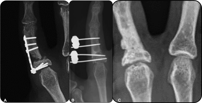

In addition, we performed an arthrolysis, which partially improved mobility (intraoperative MCP arch: 0° to 70°), and early mobilization was started within 48 hours.
After surgery, the patient presented progressive pain relief, with a visual analog scale (VAS) score of 4 at 3 months, and of 0 at 6 months; in addition, the CT and MRI scans showed recovery of joint morphology ([Figure 9]) from an initial 21 × 18 mm defect to full joint coverage.

Patient 2
A 54-year-old male patient who presented with a wrist with stage-II scapholunate advanced collapse (SLAC) underwent a lunate-capitate arthrodesis. Osteosynthesis material migration resulted in a protrusion at the radiocarpal level, with chondral injury and pain ([Figures 10A,B]).

The AMIC joint salvage technique was used at the radial and lunate surfaces to avoid a radiocarpal arthrodesis ([Figures 11A,B]). Joint mobilization without resistance was started 6 days after the intervention, when the patient tolerated the pain. A protection splint was used overnight for 3 weeks. Flexion and extension improved by 20° and 30° respectively; pain relief occurred almost immediately, and it was sustained over time ([Figures 12A,B]). The last follow-up, at 5 years, revealed no pain and preserved function, and CT scans at 2, 4, and 6 months confirmed joint recovery ([Figures 13A,B,C]) with adequate coverage of the initial radial (16 × 6 mm) and lunate (11 × 6 mm) defects.


Patient 3
A patient suffered trauma in wrist hyperextension resulting in persistent pain, and came to a consultation two months later. The CT scans showed the absence of a whole lunate chondral fragment ([Figures 14A,B,C]).

Through an arthroscopic approach, the superficial tissue was excised, debrided, and perforated; next, the defect was filled with a cancellous distal radial bone graft compacted with Tissucol ([Figures 15A,B]) to level the osteochondral defect to the articular surface of the remaining lunate bone, followed by membrane coverage ([Figure 15C]).

Active mobilization with no resistance was allowed seven days after surgery given the stablility of the matrix implant. Currently, 5.5 years after the procedure, the patient remains asymptomatic.
Other cases
This technique has also been used to treat Badia grade-II rhizarthrosis ([Figures 16A,B,C]), posttraumatic chondral defects in distal radius fractures ([Figures 17A,B,C]), and sequelae from Bennett fracture-dislocations, or fractures at the base of the first phalange or at the head of the MTC bone.


Therefore, its indication encompasses any joint with an irrecoverable chondral injury whose surgical alternative is palliative surgery, either arthrodesis, proximal carpectomy, arthroplasty etc.
It is especially important to perserve the anatomy of the TMC joint without altering the saddle shape of the trapezius. One should also be meticulous when lowering the surface to adapt it to the thickness of the membrane for an easier adaptation of small fragments instead of a single piece.
Conclusions
The AMIC technique has proven to be an effective alternative to treat chondral lesions in other joints.
It is more successful than microfractures or nanofractures used in isolation, and much less expensive and complex than treatments based on chondrocyte culture.
We still need to determine if these good outcomes can be extrapolated to a small joint. Adequately planned studies could enable us to obtain statistically significant findings that scientifically support the good first impressions with the AMIC technique in these hand and wrist chondral injuries.
Conflict of Interests
The authors have no conflict of interests to declare.
-
References
- 1 Benthien JP, Behrens P. The treatment of chondral and osteochondral defects of the knee with autologous matrix-induced chondrogenesis (AMIC): method description and recent developments. Knee Surg Sports Traumatol Arthrosc 2011; 19 (08) 1316-1319
- 2 Piontek T, Ciemniewska-Gorzela K, Szulc A, Naczk J, Słomczykowski M. All-arthroscopic AMIC procedure for repair of cartilage defects of the knee. Knee Surg Sports Traumatol Arthrosc 2012; 20 (05) 922-925
- 3 Behrens P. Matrixgekoppelte Mikrofrakturierung. Ein neues Konzept zur Knorpeldefektbehandlung. Arthroskopie 2005; 18 (03) 193-197
- 4 Pridie K. A method of resurfacing osteoarthritic knee joints. J Bone Joint Surg [Br] 5 1959; 41: 618-619
- 5 Steadman JR, Rodkey WG, Rodrigo JJ. Microfracture: surgical technique and rehabilitation to treat chondral defects. Clin Orthop Relat Res 2001; (391, Suppl) S362-S369
- 6 Lee KB, Bai LB, Chung JY, Seon JK. Arthroscopic microfracture for osteochondral lesions of the talus. Knee Surg Sports Traumatol Arthrosc 2010; 18 (02) 247-253
- 7 Chuckpaiwong B, Berkson EM, Theodore GH. Microfracture for osteochondral lesions of the ankle: outcome analysis and outcome predictors of 105 cases. Arthroscopy 2008; 24 (01) 106-112
- 8 Hannon CP, Murawski CD, Fansa AM, Smyth NA, Do H, Kennedy JG. Microfracture for osteochondral lesions of the talus: a systematic review of reporting of outcome data. Am J Sports Med 2013; 41 (03) 689-695
- 9 Gobbi A, Karnatzikos G, Kumar A. Long-term results after microfracture treatment for full-thickness knee chondral lesions in athletes. Knee Surg Sports Traumatol Arthrosc 2014; 22 (09) 1986-1996
- 10 Benthien JP, Behrens P. Nanofractured autologous matrix induced chondrogenesis (NAMIC©)–Further development of collagen membrane aided chondrogenesis combined with subchondral needling: A technical note. Knee 2015; 22 (05) 411-415
- 11 Peñalver JM, Villalba J, Yela-Verdú CP. et al. All-Arthroscopic Nanofractured Autologous Matrix-Induced Chondrogenesis (A-NAMIC) Technique for the Treatment of Focal Chondral Lesions of the Knee. Arthrosc Tech 2020; 9 (06) e755-e759
- 12 Migliorini F, Eschweiler J, Maffulli N. et al. Autologous Matrix Induced Chondrogenesis (AMIC) Compared to Microfractures for Chondral Defects of the Talar Shoulder: A Five-Year Follow-Up Prospective Cohort Study. Life (Basel) 2021; 11 (03) 244 Pages 1–9 .
- 13 Valderrabano V, Miska M, Leumann A, Wiewiorski M. Reconstruction of Osteochondral Lesions of the Talus With Autologous Spongiosa Grafts and Autologous Matrix-Induced Chondrogenesis. The American Journal of Sports Medicine 2013; 41 (03) 519-527
- 14 Steinwachs M, Kreuz PC. Autologous Chondrocyte Implantation in Chondral Defects of the Knee With a Type I/III Collagen Membrane: A Prospective Study With a 3-Year Follow-up. Arthroscopy: The Journal of Arthroscopic & Related Surgery 2007; 23 (04) 381-387
- 15 Benthien JP, Behrens P. Autologous matrix-induced chondrogenesis (AMIC). A one-step procedure for retropatellar articular resurfacing. Acta Orthop Belg 2010; 76 (02) 260-263
- 16 Steinwachs MR, Gille J, Volz M, Anders S, Jakob R, De Girolamo L, Wittmann U. Systematic Review and Meta-Analysis of the Clinical Evidence on the Use of Autologous Matrix-Induced Chondrogenesis in the Knee. 2019. 1-15 CARTILAGE 194760351987084. doi:10.1177/1947603519870846
- 17 Gao L, Orth P, Cucchiarini M, Madry H. Autologous Matrix-Induced Chondrogenesis: A Systematic Review of the Clinical Evidence. Am J Sports Med 2019; Jan; 47 (01) 222-231
- 18 Becher C, Malahias MA, Ali MM, Maffulli N, Thermann H. (2018). Arthroscopic microfracture vs. arthroscopic autologous matrix-induced chondrogenesis for the treatment of articular cartilage defects of the talus. Knee Surgery, Sports Traumatology, Arthroscopy 2019; 27: 2731-2736
- 19 Kaufman D, Etcheson J, Yao J. Microfracture for Ulnar Impaction Syndrome: Surgical Technique and Outcomes with Minimum 2-Year Follow-up. Journal of Wrist Surgery 2016; 6 (01) 60-64
- 20 Kaiser N, Jacobi M, Kusano T. et al. Clinical results 10 years after AMIC in the knee. Swiss Med Wkly 2015; 145 (Suppl. 210) 43S
- 21 Gille J, Kunow J, Boisch L. et al. Cell-Laden and Cell-Free Matrix-Induced Chondrogenesis versus Microfracture for the Treatment of Articular Cartilage Defects: A Histological and Biomechanical Study in Sheep. Cartilage 2010; 1 (01) 29-42
- 22 Volz M, Schaumburger J, Frick H, Grifka J, Anders S. A randomized controlled trial demonstrating sustained benefit of Autologous Matrix-Induced Chondrogenesis over microfracture at five years. Int Orthop 2017; 41 (04) 797-804
- 23 Kramer J, Böhrnsen F, Lindner U. et al. In vivo matrix-guided human mesenchymal stem cells. Cell Mol Life Sci 2006; 63 (05) 616-626
- 24 Gomoll AH, Farr J, Gillogly SD. et al. Surgical management of articular cartilage defects of the knee. J Bone Joint Surg Am 2010; 92 (14) 2470-2490
- 25 Mumme M, Barbero A, Miot S. et al. Nasal chondrocyte-based engineered autologous cartilage tissue for repair of articular cartilage defects: an observational first-in-human trial. Lancet 2016; 388 (10055): 1985-1994
- 26 Fulco I, Miot S, Haug MD. et al. Engineered autologous cartilage tissue for nasal reconstruction after tumour resection: an observational first-in-human trial. Lancet 2014; 384 (9940): 337-346
Address for correspondence
Publikationsverlauf
Eingereicht: 24. August 2021
Angenommen: 01. Oktober 2021
Artikel online veröffentlicht:
13. Dezember 2021
© 2021. SECMA Foundation. This is an open access article published by Thieme under the terms of the Creative Commons Attribution-NonDerivative-NonCommercial License, permitting copying and reproduction so long as the original work is given appropriate credit. Contents may not be used for commecial purposes, or adapted, remixed, transformed or built upon. (https://creativecommons.org/licenses/by-nc-nd/4.0/)
Thieme Revinter Publicações Ltda.
Rua do Matoso 170, Rio de Janeiro, RJ, CEP 20270-135, Brazil
-
References
- 1 Benthien JP, Behrens P. The treatment of chondral and osteochondral defects of the knee with autologous matrix-induced chondrogenesis (AMIC): method description and recent developments. Knee Surg Sports Traumatol Arthrosc 2011; 19 (08) 1316-1319
- 2 Piontek T, Ciemniewska-Gorzela K, Szulc A, Naczk J, Słomczykowski M. All-arthroscopic AMIC procedure for repair of cartilage defects of the knee. Knee Surg Sports Traumatol Arthrosc 2012; 20 (05) 922-925
- 3 Behrens P. Matrixgekoppelte Mikrofrakturierung. Ein neues Konzept zur Knorpeldefektbehandlung. Arthroskopie 2005; 18 (03) 193-197
- 4 Pridie K. A method of resurfacing osteoarthritic knee joints. J Bone Joint Surg [Br] 5 1959; 41: 618-619
- 5 Steadman JR, Rodkey WG, Rodrigo JJ. Microfracture: surgical technique and rehabilitation to treat chondral defects. Clin Orthop Relat Res 2001; (391, Suppl) S362-S369
- 6 Lee KB, Bai LB, Chung JY, Seon JK. Arthroscopic microfracture for osteochondral lesions of the talus. Knee Surg Sports Traumatol Arthrosc 2010; 18 (02) 247-253
- 7 Chuckpaiwong B, Berkson EM, Theodore GH. Microfracture for osteochondral lesions of the ankle: outcome analysis and outcome predictors of 105 cases. Arthroscopy 2008; 24 (01) 106-112
- 8 Hannon CP, Murawski CD, Fansa AM, Smyth NA, Do H, Kennedy JG. Microfracture for osteochondral lesions of the talus: a systematic review of reporting of outcome data. Am J Sports Med 2013; 41 (03) 689-695
- 9 Gobbi A, Karnatzikos G, Kumar A. Long-term results after microfracture treatment for full-thickness knee chondral lesions in athletes. Knee Surg Sports Traumatol Arthrosc 2014; 22 (09) 1986-1996
- 10 Benthien JP, Behrens P. Nanofractured autologous matrix induced chondrogenesis (NAMIC©)–Further development of collagen membrane aided chondrogenesis combined with subchondral needling: A technical note. Knee 2015; 22 (05) 411-415
- 11 Peñalver JM, Villalba J, Yela-Verdú CP. et al. All-Arthroscopic Nanofractured Autologous Matrix-Induced Chondrogenesis (A-NAMIC) Technique for the Treatment of Focal Chondral Lesions of the Knee. Arthrosc Tech 2020; 9 (06) e755-e759
- 12 Migliorini F, Eschweiler J, Maffulli N. et al. Autologous Matrix Induced Chondrogenesis (AMIC) Compared to Microfractures for Chondral Defects of the Talar Shoulder: A Five-Year Follow-Up Prospective Cohort Study. Life (Basel) 2021; 11 (03) 244 Pages 1–9 .
- 13 Valderrabano V, Miska M, Leumann A, Wiewiorski M. Reconstruction of Osteochondral Lesions of the Talus With Autologous Spongiosa Grafts and Autologous Matrix-Induced Chondrogenesis. The American Journal of Sports Medicine 2013; 41 (03) 519-527
- 14 Steinwachs M, Kreuz PC. Autologous Chondrocyte Implantation in Chondral Defects of the Knee With a Type I/III Collagen Membrane: A Prospective Study With a 3-Year Follow-up. Arthroscopy: The Journal of Arthroscopic & Related Surgery 2007; 23 (04) 381-387
- 15 Benthien JP, Behrens P. Autologous matrix-induced chondrogenesis (AMIC). A one-step procedure for retropatellar articular resurfacing. Acta Orthop Belg 2010; 76 (02) 260-263
- 16 Steinwachs MR, Gille J, Volz M, Anders S, Jakob R, De Girolamo L, Wittmann U. Systematic Review and Meta-Analysis of the Clinical Evidence on the Use of Autologous Matrix-Induced Chondrogenesis in the Knee. 2019. 1-15 CARTILAGE 194760351987084. doi:10.1177/1947603519870846
- 17 Gao L, Orth P, Cucchiarini M, Madry H. Autologous Matrix-Induced Chondrogenesis: A Systematic Review of the Clinical Evidence. Am J Sports Med 2019; Jan; 47 (01) 222-231
- 18 Becher C, Malahias MA, Ali MM, Maffulli N, Thermann H. (2018). Arthroscopic microfracture vs. arthroscopic autologous matrix-induced chondrogenesis for the treatment of articular cartilage defects of the talus. Knee Surgery, Sports Traumatology, Arthroscopy 2019; 27: 2731-2736
- 19 Kaufman D, Etcheson J, Yao J. Microfracture for Ulnar Impaction Syndrome: Surgical Technique and Outcomes with Minimum 2-Year Follow-up. Journal of Wrist Surgery 2016; 6 (01) 60-64
- 20 Kaiser N, Jacobi M, Kusano T. et al. Clinical results 10 years after AMIC in the knee. Swiss Med Wkly 2015; 145 (Suppl. 210) 43S
- 21 Gille J, Kunow J, Boisch L. et al. Cell-Laden and Cell-Free Matrix-Induced Chondrogenesis versus Microfracture for the Treatment of Articular Cartilage Defects: A Histological and Biomechanical Study in Sheep. Cartilage 2010; 1 (01) 29-42
- 22 Volz M, Schaumburger J, Frick H, Grifka J, Anders S. A randomized controlled trial demonstrating sustained benefit of Autologous Matrix-Induced Chondrogenesis over microfracture at five years. Int Orthop 2017; 41 (04) 797-804
- 23 Kramer J, Böhrnsen F, Lindner U. et al. In vivo matrix-guided human mesenchymal stem cells. Cell Mol Life Sci 2006; 63 (05) 616-626
- 24 Gomoll AH, Farr J, Gillogly SD. et al. Surgical management of articular cartilage defects of the knee. J Bone Joint Surg Am 2010; 92 (14) 2470-2490
- 25 Mumme M, Barbero A, Miot S. et al. Nasal chondrocyte-based engineered autologous cartilage tissue for repair of articular cartilage defects: an observational first-in-human trial. Lancet 2016; 388 (10055): 1985-1994
- 26 Fulco I, Miot S, Haug MD. et al. Engineered autologous cartilage tissue for nasal reconstruction after tumour resection: an observational first-in-human trial. Lancet 2014; 384 (9940): 337-346


