Keywords
lipoatrophy - lupus panniculitis - fat grafting - autologous fat transfer
Introduction
Cutaneous lupus erythematosus (CLE) has a variety of manifestations, which can be
divided into three categories: acute cutaneous lupus erythematosus (ACLE), subacute
cutaneous lupus erythematosus (SCLE), and chronic cutaneous lupus erythematosus (CCLE).
Lupus erythematosus panniculitis or lupus panniculitis (LP), which is a variant of
CCLE, occurs from inflammation of the subcutaneous fat and leads to tender nodules
and ulcerative lesions with a chronic relapse nature. Even though various kinds of
medical treatment have been applied, LP often causes scarring and postinflammatory
lipoatrophy with marked contour deficits.[1]
[2]
Up to the present date, the current mainstay treatment for CLE is medical treatment,
including topical corticosteroids and calcineurin inhibitors, systemic antimalarials,
and systemic steroids. Other immunomodulators are considered for refractory disease
or in cases of contraindication in systemic steroid usage.[3]
The treatment of atrophic cutaneous lupus lesion by injectables is avoided due to
the theoretical risk of disease reactivation by tissue trauma.[4] Indeed, a previous study involved the injection of hyaluronic acid and poly-L-lactic
acid for the treatment of LP-induced facial lipoatrophy and found that the effect
of the treatment was only temporary and the reactivation of disease was suspected
from the injected materials.[5] However, the data from several studies have demonstrated satisfactory outcomes and
favorable safety profile of fat grafting in LP lesions.[6]
[7]
Autologous fat transfer, also known as fat grafting, has been used for volume restoration
and contour defects in reconstructive surgery for decades. Autologous fat grafting
not only involves volume replacement but also has the ability to regenerate itself.
Several studies have demonstrated the presence of multipotent stem cells in the stromal
vascular fraction of processed fat grafts, which can promote angiogenesis, alter the
apoptosis process, and modulate immune responses.[4]
[5]
[6]
[7]
[8]
The keystone of potential treatment is the use of adipose-derived stem cells (ADSCs),
that are capable of soft tissue regeneration and of restoring devitalized tissue,
as seen in many studies, including wound healing promotion, antiaging treatment, and
damaged skin rejuvenation.[9]
[10] In this study, we aim to share our experience regarding the treatment of lipoatrophy
secondary to LP with autologous fat transfer.
Case
A 48-year-old female presented with an atrophic lesion at her left temporal area for
2 years. The lesion was first occurred with erythematous plaque, tender nodules, and
intermittent ulcers followed by depression of the lesion, which was progressive for
a year then stabilized. The patient also had a rash at her left cheek, which was an
ill-defined indurated erythematous plaque with sparing of the nasolabial fold. Both
the rashes were aggravated by sunlight exposure and heat. The patient denied any underlying
disease, current medications, or history of photosensitivity rash in other areas.
Physical examination showed locally subcutaneous atrophic lesion at the left temporal
region and an ill-defined indurated erythematous plaque at the left cheek with sparing
of the nasolabial fold, while no facial palsy, no bone deformity at the temporal area,
no malocclusion, no other skin lesion, no arthritis, no oral ulcer, and no alopecia
were found ([Figs. 1A] and [2A]).
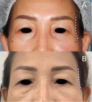 Fig. 1 The patient had an ill-defined erythematous plaque with local subcutaneous atrophy
at the left temporal region and an ill-defined indurated erythematous plaque at the
left cheek at the first visit (A). At 1 year after autologous fat transfer, the patient showed an improvement in the
volume deficit at the left temporal region (B). All photographs were consented for publish by the patient.
Fig. 1 The patient had an ill-defined erythematous plaque with local subcutaneous atrophy
at the left temporal region and an ill-defined indurated erythematous plaque at the
left cheek at the first visit (A). At 1 year after autologous fat transfer, the patient showed an improvement in the
volume deficit at the left temporal region (B). All photographs were consented for publish by the patient.
 Fig. 2 The ill-defined erythematous plaque with local subcutaneous atrophy at the left temporal
region before treatment (A). Improvements in the left temporal volume depletion and erythematous plaque together
with the indurated plaque at the left cheek after 1 year of the first autologous fat
transfer and medical treatment (B). All photographs were consented for publish by the patient.
Fig. 2 The ill-defined erythematous plaque with local subcutaneous atrophy at the left temporal
region before treatment (A). Improvements in the left temporal volume depletion and erythematous plaque together
with the indurated plaque at the left cheek after 1 year of the first autologous fat
transfer and medical treatment (B). All photographs were consented for publish by the patient.
The patient was sent to a rheumatologist and dermatologist for evaluation. Autoimmune
panel tests were done. The patient had a positive antinuclear antibody with a fine-speckled
pattern titer 1:320, positive anti-cytoplasmic antibody titer 1:100, and positive
anti-Ro autoantibodies 2 + , while the other antibody tests were unremarkable and
there was no evidence of internal organ involvement. The diagnoses were tumid lupus
erythematosus (tumid LE) for the indurated lesion at the cheek and LP with secondary
lipoatrophy for the lesion at the left temporal area.
Unfortunately, the patient was unwilling to have a skin biopsy performed, which limited
the confirmation of CLE pathologically. However, the erythematous plaque responded
well to treatment with oral hydroxychloroquine 200 mg once daily and topical 0.1%
mometasone furoate cream. The medical treatment had been continued for 10 months before
the autologous fat grafting. The patient was scheduled for operation after the disease
was stabilized by medications as mentioned above to prevent the secondary damage to
the donor site. None of surgical treatment and injectables was applied to the patient
prior autologous fat grafting.
For the lipoatrophy at the left temporal area, the volume of fat grafting overcorrected
the lesion volume for 20 to 30% excess to achieve the favorable outcome after fat
resorption. The patient underwent autologous 8 mL fat transfer injection at the left
temporal area. The fat grafting was harvested from a subcutaneous layer of the lower
abdominal area using the tumescent technique and no donor site morbidity was reported.
At 2 months follow-up, the patient reported an improvement in the previously treated
lesion at temporal area with a minimal volume loss of grafted fat. Furthermore, the
tender nodule and intermittent ulcers were also resolved.
After 1 year of antimalarial and topical corticosteroids, the erythematous plaque
at the left cheek and left temporal area was completely resolved. However, a depressed
contour of left temporal area was still noted, and the patient asked for the second
fat grafting procedure. The patient underwent the second session at a 16-month interval
due to fat resorption from the hydroxychloroquine. She received 3.7 mL of autologous
fat injection, which was harvested from subcutaneous fat of the lower abdomen with
the tumescent technique. Neither an adverse event from the procedure nor reactivation
of the disease was reported and the patient was satisfied with the volume restoration
of the atrophic lesion and remission of the tender nodule and intermittent ulcers
at the temporal area ([Figs. 1B] and [2B]).
Discussion
Unlike other forms of CLE, LP commonly disturbs the patient not only from the inflammation
during the active phase of disease but also from the scarring and lipoatrophy after
inflammation subsides. Secondary lipoatrophy from LP can affect the appearance and
contour of the patient's skin, which can lead to a disturbance of the patient's self-esteem
and quality of life.
The current mainstay for CLE treatment is medical treatment. However, injectable treatment
has been avoided as it carries a theoretical risk of disease reactivation by tissue
trauma.[4] The study of volume restoration in LP associated-lipoatrophy by dermal fillers provided
satisfied results but there were risks of disease exacerbation and a granulomatous
reaction from the antigenicity of the injectables.[5]
The treatment of LP with injectables should be avoided in active inflammatory phase
since the LP can be exacerbated by trauma. Adequate medical treatment to control the
disease activation before fat grafting is important.[4]
[5] So, the autologous fat grafting should be done in the stable and non-inflammatory
phase of disease.
In this report, the patient received autologous fat transfer for volume restoration
of a lipoatrophic lesion. The results were promising and the patient was satisfied
with the outcome. The patient reported that the reactivation of the disease, which
involved the spontaneous presence of tender nodules and intermittent ulcers, was gradually
resolved after the first autologous fat transfer treatment. The patient was satisfied
with the improvement in her disfigurement and the endurance of the fat grafting for
over a year. Hence, autologous fat grafting should be considered as a safe and simple
procedure with a positive long-lasting effect. However, a staged procedure might be
required to achieve the target volume since the volume depletion of injected fat.
The functions of ADSCs has been proposed in several literatures such as promotion
of angiogenesis, alteration the apoptosis process, and modulation of immune responses.[8]
[9]
[10] Apart from the direct volume correction by fat grafting injection, we observed the
disease stabilization of the intermittent ulcer at LP lesion after fat grafting injection.
The quiescence of LP lesion might have resulted from immunomodulatory effects of the
ADSCs, which suppress the inflammatory process of LP.
In summary, the treatment of secondary lipoatrophy from LP with autologous fat grafting
shows promising long-lasting results in volume restoration and can alleviate spontaneous
reactivation of the LP lesion without adverse events. This treatment should be done
in the stable and quiescent phase of the disease.


