Introduction
The late effects of adjuvant radiation therapy (RT) encompass a spectrum of manifestations,
including hair loss, pigmentary changes, loss of flap volume, and fibrosis, which
appear from 6 months and continue till several years postradiotherapy.[1] These persistent changes occur due to radiation-induced tissue hypoxia, attributed
to capillary endothelial damage by ionizing radiation.[2] This problem is especially relevant in the context of head and neck reconstruction,
as transferred tissue flaps frequently experience significant soft tissue fibrosis
after RT which negatively affects both their functionality and appearance.[3]
[4]
[5] Tissue hypoxia, which is a major factor in causing these long-term issues, can worsen
due to subclinical infections caused by minor injuries, exposed implants, or the presence
of underlying osteoradionecrosis.[2] The cumulative impact of these factors can lead to significant soft tissue deformities
resulting in functional and aesthetic compromise. In certain cases, replacement of
affected tissue by another soft tissue flap mitigates the late complication of adjuvant
RT.
A series of 21 patients who required replacement of previously transferred flaps with
a second composite tissue transfer for secondary soft tissue changes following adjuvant
RT is presented.
Materials and Methods
Retrospective data from January 2019 to 2023, retrieved from electronic medical records,
were analyzed. Of 756 patients who had undergone primary excision and reconstruction,
21 individuals underwent a secondary soft tissue transfer to address severe soft tissue
changes related to adjuvant RT. The age group of the patients ranged from 29 to 70
years, of these 17 were males and 5 were females.
Patients with tumor recurrence, plate removal alone, without soft tissue transfer,
or those who underwent minor local tissue readjustments without soft tissue transfers
were excluded. Those who underwent supplementary secondary procedures like fat grafting
and scar revisions were likewise omitted from the analysis.
The parameters recorded included the clinical presentation and soft tissue fibrosis
along with its underlying pathology requiring surgery. Volume loss ([Fig. 1]), scarring ([Fig. 2]), exposed hardware or fistulas leading to functional impairment ([Figs. 3] and [4]), the duration elapsed since completion of radiotherapy, and surgical method employed
(free tissue transfer or pedicled flap cover) were recorded. Recipient vessels chosen
and the ultimate outcome (focused on whether the intervention successfully achieved
its intended goal) were also documented ([Table 1]).
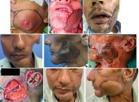 Fig. 1 (A) Primary marking showing extent of full-thickness excision. (B) Primary reconstruction with Fibula osseocutaneous free flap (FOCFF) and Anterolateral
thigh (ALT) free flap. (C) Postsurgery prior to radiotherapy. (D) Severe soft tissue fibrosis with leathery, pigmented, and contracted skin along
with discharging sinus and significant volume loss. (E) Topographic markings to show the extent of volumetric replacement planned using
a differentially thinned flap (ALT). (F) Prior identification of superficial temporal vessels. (G) Volume and contour restored after secondary soft tissue transfer (ALT).
Fig. 1 (A) Primary marking showing extent of full-thickness excision. (B) Primary reconstruction with Fibula osseocutaneous free flap (FOCFF) and Anterolateral
thigh (ALT) free flap. (C) Postsurgery prior to radiotherapy. (D) Severe soft tissue fibrosis with leathery, pigmented, and contracted skin along
with discharging sinus and significant volume loss. (E) Topographic markings to show the extent of volumetric replacement planned using
a differentially thinned flap (ALT). (F) Prior identification of superficial temporal vessels. (G) Volume and contour restored after secondary soft tissue transfer (ALT).
 Fig. 2 (A) Postresection of carcinoma upper alveolus and nasal floor. (B) Primary reconstruction with Deep circumflex iliac artery (DCIA) and Radial artery
forearm flap (RAFF) (note extent of overcorrection of lip). (C) Seven months postradiotherapy. (D) Two and half year postadjuvant radiotherapy showing complete loss of volume, causing
deformity and incompetence of the upper lip. (E) Secondary soft tissue transfer with RAFF, to restore the lip along with placement
of dental implants.
Fig. 2 (A) Postresection of carcinoma upper alveolus and nasal floor. (B) Primary reconstruction with Deep circumflex iliac artery (DCIA) and Radial artery
forearm flap (RAFF) (note extent of overcorrection of lip). (C) Seven months postradiotherapy. (D) Two and half year postadjuvant radiotherapy showing complete loss of volume, causing
deformity and incompetence of the upper lip. (E) Secondary soft tissue transfer with RAFF, to restore the lip along with placement
of dental implants.
 Fig. 3 (A) Carcinoma lower lip showing extent of excision. (B) Primary reconstruction with Radial artery forearm flap (RAFF). (C) Six months postadjuvant radiation therapy —volume loss, exposed gingiva, loss of
lip competence, and drooling. (D) One year postadjuvant RT—showing progress of soft tissue fibrosis. (E) Secondary soft tissue transfer (RAFF) with restoration of volume and lip competence.
Fig. 3 (A) Carcinoma lower lip showing extent of excision. (B) Primary reconstruction with Radial artery forearm flap (RAFF). (C) Six months postadjuvant radiation therapy —volume loss, exposed gingiva, loss of
lip competence, and drooling. (D) One year postadjuvant RT—showing progress of soft tissue fibrosis. (E) Secondary soft tissue transfer (RAFF) with restoration of volume and lip competence.
 Fig. 4 (A) Defect postexcision for carcinoma buccal mucosa. (B) Primary reconstruction with chimeric fibula osseocutaneous free flap (FOCFF) and
proximal peroneal artery flap restoring adequate volume. (C) Immediately post-radiation therapy showing acute changes. (D) Osteoradionecrosis (ORN) with orocutaneous fistula along with severe soft tissue
fibrosis. (E) Postreconstruction with double island Radial artery forearm flap (RAFF).
Fig. 4 (A) Defect postexcision for carcinoma buccal mucosa. (B) Primary reconstruction with chimeric fibula osseocutaneous free flap (FOCFF) and
proximal peroneal artery flap restoring adequate volume. (C) Immediately post-radiation therapy showing acute changes. (D) Osteoradionecrosis (ORN) with orocutaneous fistula along with severe soft tissue
fibrosis. (E) Postreconstruction with double island Radial artery forearm flap (RAFF).
Table 1
Master char
|
No.
|
Age/Sex
|
Diagnosis
|
Primary reconstruction
|
Neck dissection
|
RT dose (Gy)
|
Indication for secondary soft tissue transfer
|
Secondary flap used
|
Time between adjuvant RT and secondary reconstruction (mo)
|
Recipient vessels A/V
|
Follow-up
(mo)
|
|
1
|
60/M
|
CA BM
|
PMMC
|
Unilateral
|
60
|
ORN
|
RAFF
|
13
|
Contralateral FA and IJV
|
66
|
|
2
|
50/M
|
CA BM
|
FOCFF
|
Unilateral
|
60
|
Volume loss and contour irregularity
|
ALT
|
24
|
Ipsilateral FA and EJV
|
65
|
|
3
|
43/M
|
CA BM
|
FOCFF
|
Bilateral
|
60
|
Exposed implant, volume loss, and contour irregularity
|
RAFF
|
12
|
Contralateral FA and EJV
|
60
|
|
4
|
39/M
|
CA BM
|
FOCFF
|
Unilateral
|
60
|
Exposed implant with volume loss
|
RAFF
|
22
|
Contralateral FA, IJV, and EJV
|
52
|
|
5
|
47/M
|
CA lower alveolus
|
FOCFF
|
Unilateral
|
63
|
Exposed implant with contracted skin
|
DP
|
10
|
|
41
|
|
6
|
50/M
|
CA lower alveolus
|
FOCFF
|
Unilateral
|
60
|
Exposed implant with contracted skin
|
PMMC
|
6
|
|
40
|
|
7
|
62/M
|
CA lower alveolus
|
FOCFF
|
Bilateral
|
60
|
Exposed implant with volume loss
|
RAFF
|
84
|
STA and STV
|
31
|
|
8
|
71/M
|
CA upper alveolus
|
FOCFF
|
Bilateral
|
60
|
Exposed implant with contour irregularity
|
LD
|
14
|
|
36
|
|
9
|
47/M
|
CA BM
|
FOCFF
|
Unilateral
|
60
|
Exposed implant with contour irregularity
|
PMMC
|
27
|
|
32
|
|
10[a]
|
46/F
|
CA lower lip
|
RAFF
|
Bilateral
|
60
|
Volume loss with lower lip incompetence
|
RAFF
|
11
|
STA and STV, EJV
|
25
|
|
11
|
48/F
|
CA lower alveolus
|
FOCFF + RAFF
|
Unilateral
|
60
|
Exposed implant with orocutaneous fistula
|
DP flap
|
5
|
|
23
|
|
12
|
57/M
|
CA lower alveolus
|
FOCFF
|
Bilateral
|
60
|
Exposed Implant, fibrosed skin
|
RAFF
|
13
|
STA/STV and EJV
|
18
|
|
13
|
60/M
|
CA BM
|
FOCFF
|
Unilateral
|
60
|
Exposed implant with discharging sinus
|
DP Flap
|
10
|
|
6
|
|
14
|
55/M
|
CA lower alveolus
|
RAFF + DCIA
|
Unilateral
|
60
|
Fistula with fibrosed skin
|
ALT
|
15
|
STA and STV
|
14
|
|
15[a]
|
41/M
|
CA lower alveolus
|
FOCFF + ALT
|
Unilateral
|
60
|
Exposed implant, contour irregularity
|
ALT
|
75
|
STA and 2 STV
|
3
|
|
16[a]
|
72/F
|
CA lower alveolus
|
FOCFF
|
Bilateral
|
60
|
Orocutaneous fistula with ORN
|
RAFF
|
105
|
STA and STV, EJV
|
5
|
|
17
|
57/M
|
CA central upper alveolus
|
DCIA
|
Bilateral
|
60
|
Oronasal fistula, volume loss, and contour irregularity
|
RAFF
|
11
|
STA and STV, EJV
|
2
|
|
18[a]
|
55/M
|
CA central upper alveolus
|
RAFF + DCIA
|
Unilateral
|
60
|
Volume loss and contour irregularity with incompetent upper lip
|
RAFF
|
30
|
STA and STV
|
1
|
|
19
|
42/M
|
CA lower alveolus
|
FOCFF
|
Bilateral
|
60
|
Flap volume loss, contour irregularity, and orocutaneous fistula
|
ALT
|
14
|
STA and STV
|
1
|
|
20
|
29/F
|
CA maxilla
|
Free LD
|
Unilateral
|
60
|
Exposed implant with contour irregularity
|
Forehead
|
8
|
|
4
|
|
21
|
60/M
|
CA lower alveolus
|
FOCFF
|
Bilateral
|
|
Exposed implant with orocutaneous fistula
|
RAFF
|
38
|
STA and STV
|
1
|
Abbreviations: ALT, anterolateral thigh flap; BM, buccal mucosa; CA, Carcinoma; DCIA,
deep circumflex iliac artery flap; DP, deltopectoral flap; EJV, external jugular vein;
F, female; FA, facial artery; FOCFF, fibula osseocutaneous free flap; IJV, internal
jugular vein; LD, latissimus dorsi flap; M, male; ORN, osteoradionecrosis; PMMC, pectoralis
major myocutaneous flap; RAFF, radial artery forearm flap; RT, radiation therapy;
STA, superficial temporal artery; STV, superficial temporal vein.
a Illustrated cases.
Results
The age demography of the cohort ranged from 29 to 70 years of which 17 were males
and 5 were females.
Primarily 10 patients had complex through-and-through defects, of these 4 were reconstructed
using double free flaps ([Table 1]) ([Figs. 1] and [4]). Chimeric, fibula osseocutaneous flap, combined with proximal peroneal artery perforator
flap were used in five of those patients and a radial artery forearm flap was used
for lining and cover in one patient. The other 11 patients were addressed using single
flaps ([Table 1]).
All patients had received external beam radiation, using intensity-modulated RT (IMRT)
with photon beams, delivering a total of 60 Gy over 30 fractions to the tumor (flap)
bed. The period, from the conclusion of adjuvant radiotherapy and surgical intervention
ranged from 5 to 108 months, with a mean of 20 months.
Nine patients presented with a discharging sinus with or without exposed plate, while
8 patients presented with exposed implant. Two patients had orocutaneous fistula ([Fig. 4]), two patients complained of drooling and exposed gingiva with loss of lip competence
([Fig. 3]), and two patients presented with deformity ([Fig. 2]). Clinically associated with this underlying cause, the previously transferred flap
was found to be pigmented, leathery, oedematous, and densely scarred ([Fig. 1]).
Out of these 21 patients, 14 underwent a second free tissue transfer and 7 locoregional
tissue cover. Of the 14 microvascular tissue transplants, radial artery flap was employed
for 10, while the anterolateral thigh flap was used for 4 patients. Among the 7 regional
flaps that were transferred, the deltopectoral flap (DP) was the most frequent, for
3 patients, followed by the pectoralis major myocutaneous flap for 2, while the latissimus
dorsi myocutaneous flap and paramedian forehead flap were utilized for one patient
each ([Table 1]).
In 10 patients, the superficial temporal vessels were chosen as recipient vessels,
while the unoperated contralateral neck vessels were chosen for 3 patients. Only in
one patient the previously operated and radiated ipsilateral neck recipient vessel
was found suitable.
Two patients had follow-up beyond 3 years, while 11 patients had been followed up
for more than 6 months and 7 patients had a shorter follow-up.
Discussion
In contrast to acute post-RT changes, the late sequelae of adjuvant radiotherapy is
stated to start beyond 6 months and continue for several years.[5] The incidence is reported to be around 10 to 15%.[6]
[7]
[8] Over long term, radiated tissue flaps can experience various significant changes,
including fibrosis, volume reduction, osteoradionecrosis, plate exposure, and fistula.[4]
[5] These alterations in the flap's characteristics resulting from adjuvant RT can pose
challenges both in terms of function and appearance ([Fig. 2]).
Ionizing radiation primarily damages the deoxyribonucleic acid and alters the cellular
microenvironment through free radicals.[9] The mechanism of underlying soft tissue damage due to radiation follows the principle
that, cells with a higher rate of division are more vulnerable to radiation and suffer
more damage compared with cells not actively dividing. Among these, endothelial cells
found in arterioles and capillary networks are especially sensitive to radiation in
comparison to stromal cells. This sensitivity leads to obliterative endarteritis,
which results in reduced oxygen supply to the tissue and characteristic fibrotic changes
in the tissue's stroma that has been damaged by radiation.[2] However, in tissues with limited cell turnover, these processes are less influenced
by cell division and are instead driven by chemokines and fibrotic cytokines. This
leads to a latency period between radiation exposure and the onset of tissue damage,
including tissue fibrosis, atrophy, or vascular injury.[9] This progression is like the chronic healing process. Although various factors contribute
to the late sequelae of adjuvant radiotherapy, including treatment, patient, and tumor-related
factors, Masuda and Kamiya have highlighted that certain patients may possess a genetic
susceptibility to radiation-induced injury.[10]
Majority of late postradiation effects typically become apparent at approximately
1 year after treatment. For secondary procedures, a minimum of 6 months following
adjuvant radiotherapy is generally considered “safe” with regard to wound healing.[4] The underlying vascular endarteritis makes an attempt “to repair” growing new capillaries,
but these grow disorganized and underlying scarring and hypoxia persists.[1]
[11] In all but one of the 21 cases, the secondary procedures were performed after 6
months to as late as 10 years, following adjuvant radiotherapy.
Advancements in radiotherapy have evolved from utilizing Cobalt to photon-based techniques,
enabling precise three-dimensional dose targeting with the application of IMRT. These
innovations have indeed reduced the incidence of complications compared with earlier
methods but have not eliminated them. Patients undergoing adjuvant radiotherapy through
IMRT receive the highest radiation dose precisely focused on the excised area, which
encompasses the reconstructed flap and its surrounding region, as visualized in the
planning computed tomography. This approach ensures that a high dose is delivered
to the targeted area while significantly minimizing radiation exposure to nearby healthy
tissues.[12]
Patients typically seek medical attention only when there is a breach, discharging
sinus with exposed hardware or bone, or when fistulas develop. Patients tend to disregard
volume loss, pigmentary changes, and contour irregularities, possibly due to concerns
about additional surgical procedures, associated discomfort, and costs. Management
of plate exposure involves a conservative strategy, incorporating antibiotics and,
subsequently, plate removal, either partially or entirely. This is suitable when the
surrounding skin is pliable and can be readily closed primarily ([Algorithm 1]). However, in a specific subset of patients with plate exposure, the surrounding
soft tissue will be firm, leathery, and not pliable, making it inadequate for proper
closure. The skin might also be adherent to the underlying bone and any additional
surgical undermining of this hypoxic tissue will further compromise its vascularity
([Fig. 1]). The transfer of vascularized tissue to the radiated area offers pliable tissue
that facilitates the closure of breached areas. Moreover, it enhances volume, aesthetics,
and results in improved facial contour ([Fig. 1]).
 Algorithm 1 Algorithm for management of postradiation sequelae.
While addressing radiation-related changes an initial conservative approach may be
initiated, using antibiotics, proper nutrition, cessation of tobacco, and avoiding
any pressure and trauma[8] ([Algorithm 1]).
Hyperbaric oxygen has been found to improve tissue oxygen over a course of 30 to 40
treatments. This may stimulate angiogenesis and improve granulation, resulting in
a more elastic and less fibrotic tissue.1 This may bring about improvement in 80% but the skin in no way returns to normal.[1]
Fat grafting has been coincidentally found to improve surrounding skin quality. Cell-assisted
lipotransfer at radiated sites has been proposed.15 This may be considered for minimal volume and contour irregularity when the skin
is soft and pliable.[13] However, this approach may not be suitable when the overlying skin is fibrosed and
scarred ([Figs. 1] and [2]). Additionally, fat grafting is not effective in addressing pigmentary changes or
substantial volume replacement. Use of fat grafting to prevent secondary changes in
an irradiated bed is an area that needs exploration.[14]
Presently, it appears logical that replacement of the affected tissues, with a fresh
vascularized composite tissue, would address this problem in a select group of patients
where conservative measures fail ([Fig. 2]).
The selection of flap was customized to address specific requirements and issues unique
to each patient, particularly addressing the loss of tissue volume and color match.
The decision was also influenced by factors, including the location and availability
of suitable recipient blood vessels, the patient's preference regarding the donor
site, and cost-related considerations. In the instances of free tissue transfer radial
artery forearm flap was the choice, where volume requirement was minimal, and as it
provided a thin, pliable skin, despite the drawback of a forearm scar. In cases where
patients experienced substantial volume loss requiring additional bulk, anterolateral
thigh free flap was employed. Among the pedicle flaps, the DP flap was the preferred
option due to its color match, pliability, and cost-effectiveness, even though it
required staging. Donor site of DP flap was closed primarily, resulting in a linear
scar.
In 10 out of 21 patients (i.e., 48%), ipsilateral superficial temporal vessels served
as the preferred recipient vessels. This choice was primarily based on their location
outside the radiation field, avoiding exploring the irradiated neck. Initial surgical
step involved exploring and verifying the suitability of the superficial temporal
vessels, prior to flap harvest and transfer ([Fig. 1]). For central defects where the contralateral neck was uninvolved, it was the preferred
choice.
Wei et al have discussed second free flaps in the context of addressing complications
such as volume loss resulting from insufficient planning and issues during the primary
surgery. However, their work did not address post-RT soft tissue fibrosis.[15]
[16] It is logical that replacing a scarred hypoxic tissue with a well-vascularized tissue
will address the progressive sequelae of RT.
The impact of radiation-induced alterations in skin and subcutaneous tissue is widely
acknowledged, yet there has been a lack of objective analysis in this regard. Various
factors, including the type and dose of radiation, the patient's primary disease status,
nutritional condition, and genetic influences, can contribute to these changes.[10] While exploring primary preventive measures like overcorrecting soft tissue volume,
interposing muscle or subcutaneous fat at the reconstruction site is an avenue which
is in practice.[11] The quest to mitigate the adverse effects of radiation on soft tissue and the subsequent
demands for reconstructive surgery, represent an ongoing and complex challenge in
the field of radiation oncology and plastic surgery. Further research and clinical
exploration are imperative to develop preventive and management strategies to address
these late effects effectively and improving patient outcomes.
Conclusion
While a satisfactory reconstruction is typically accomplished during primary surgery,
the delayed consequences of adjuvant radiotherapy, particularly those involving soft
tissues, can sometimes lead to significant secondary deformities, potentially resulting
in compromised functional and aesthetic outcomes. It is important to emphasize that
not all soft tissue-related issues occurring post-adjuvant RT are a direct result
of the radiotherapy itself. Rather, a specific subset of patients is affected by these
radiation-induced effects on soft tissue. In those subsets of patients, these challenges
can be effectively managed with a secondary flap procedure. This consideration should
be integrated into the surgical treatment timeline, alongside patient counseling and
motivation.


