Keywords
PET/MRI - liver lesions - molecular imaging - multimodality scanning - neoplastic
hepatic lesions - PET/CT
Introduction
Neoplastic liver lesions present significant diagnostic challenges due to their diverse
etiology, variable morphological features, and overlapping imaging characteristics.[1] Accurate characterization and staging of these lesions are critical for guiding
optimal treatment strategies and improving patient outcomes.[2] In recent years, hybrid imaging modalities, such as positron emission tomography/magnetic
resonance imaging (PET/MRI) and PET/computed tomography (PET/CT), have emerged as
invaluable tools for the comprehensive evaluation of neoplastic liver lesions.[3]
PET/CT has become a cornerstone in oncological imaging, providing sequential metabolic
and anatomical information.[4] The fusion of functional PET data with high-resolution CT images enables precise
localization, characterization, and staging of neoplastic liver lesions, thereby facilitating
treatment planning and monitoring of therapeutic response.[5] However, soft tissue contrast of CT is not optimal for diagnosis and differentiation
of soft tissue lesions. The PET images are also affected by the number of factors
including age, blood sugar, body mass index incubation time, and hepatic steatosis
affects the liver capacity to absorb fluoro-D-glucose (FDG).
Furthermore, since PET is not a standalone modality and requires anatomical localization
and limits the detectability of small size and low contrast lesions, PET on its own
is not suitable for the detection of neoplastic hepatic lesions. Clinically significant
lesions may be missed by PET/CT due to the aforementioned limitations.
On the other hand, PET/MRI combines the metabolic insights of PET with the superior
soft tissue contrast, enhanced spatial resolution, and multiparametric imaging capabilities
of MRI.[6] This hybrid modality offers several potential advantages for the assessment of neoplastic
liver lesions, including enhanced soft tissue characterization, improved intrinsic
contrast resolution with reduced injected contrast agent, reduced radiation exposure,
and truly simultaneous acquisition providing functional, anatomical, and molecular
information.[7] In patients with renal impairment, noncontrast PET/MRI is superior to noncontrast
PET/CT.
Despite the growing interest in PET/MRI for liver imaging, its comparative efficacy
and diagnostic accuracy relative to PET/CT remain areas of ongoing investigation.[8] While PET/MRI holds promise for improving lesion detection and characterization,
challenges such as motion artifacts and limited availability hinder its widespread
clinical adoption.[9]
This study aims to provide a comparative evaluation of hybrid PET/MRI and PET/CT for
neoplastic liver lesions.[10] By synthesizing current evidence, discussing technical considerations, and evaluating
diagnostic performance, this study seeks to elucidate the strengths, limitations,
and clinical implications of each modality in liver lesion evaluation.[11] Furthermore, through a critical examination of emerging research and future directions,
this research aims to contribute to optimized diagnostic algorithms and personalized
treatment strategies for patients with neoplastic liver lesions.[12]
Materials and Methods
Place of Study
The study was conducted at Omega Hospital, Hyderabad, India.
Study Type
This study was a retrospective single-center observational cross-sectional study.
Time Period
The data of this study were collected from January 2023 to May 2024.
Study Population
This study included patients diagnosed with either primary or secondary liver neoplasms.
Among primary liver tumors, hepatocellular carcinoma (HCC) is the most common, characterized
by tumor cells resembling hepatocytes. HCC is strongly associated with chronic viral
hepatitis and cirrhosis of various origins. Other primary liver tumors in this study
include benign neoplasms associated with chronic liver parenchymal disease and cholangiocarcinoma.
Cholangiocarcinoma, a primary adenocarcinoma originating from the bile ducts, resembles
adenocarcinomas in other tissues, requiring exclusion of extrahepatic origins and
differentiation from benign biliary lesions for accurate diagnosis.
Secondary liver neoplasms encompass metastatic liver lesions, which arise from cancers
spreading to the liver from other parts of the body. The malignancies included in
this study with liver metastases are colorectal cancer, breast cancer, neuroendocrine
tumor, leiomyosarcoma of the uterus, and carcinoma vulva.
The inclusion criteria were as follows:
-
Patients of age older than 18 years with an ability to understand and hear instructions,
and remain still for ∼20 minutes (the duration of PET/MRI scan).
-
Patients with an ability to undergo a PET/MRI scan within 30 minutes after the completion
of a PET/CT scan.
-
Patients with a body weight of less than 200 kg.
The exclusion criteria were as follows:
-
Pregnant women.
-
Patients with metallic, conductive, electrically, or magnetically active implants
not labeled as MRI-safe.
-
Patients with implants labeled as MRI-unsafe.
All patients with suspected or histology-proven neoplastic liver lesions who were
referred to undergo a PET/CT scan either for diagnosis, staging, or posttreatment
staging and follow-up as treatment response evaluation, and those who passed the inclusion
criteria as stated earlier, were offered to undergo a complementary liver PET/MRI
scan by the institution. Out of 63 patients who underwent a PET/CT scan, only 18 patients
agreed to undergo a complementary PET/MRI scan. The remaining 45 patients did not
undergo a complementary PET/MRI scan due to the following reasons: claustrophobia,
additional scanning time, and inconvenience. Further, out of 18 patients who underwent
PET/MRI, only 15 were included in this study and 3 patients who underwent both PET/CT
and a complementary liver PET/MRI scan were not included in this study due to movement,
metallic artifacts, lack of cooperation, and gross ascites. The recruitment process
is summarized in [Fig. 1].
 Fig. 1 This figure shows the patient recruitment process: 63 patients who were suspected
for liver malignancy between January 2023 and May 2024 underwent PET/CT scan. Forty-five
patients who underwent PET/CT for a suspected liver malignancy did not agree to undergo
the complementary abdomen PET/MRI scan. Three patients who underwent PET/MRI scan
were excluded from the study due to movement, metallic artifacts, lack of cooperation,
and gross ascites. CT, computed tomography; MRI, magnetic resonance imaging; PET,
positron emission tomography.
Fig. 1 This figure shows the patient recruitment process: 63 patients who were suspected
for liver malignancy between January 2023 and May 2024 underwent PET/CT scan. Forty-five
patients who underwent PET/CT for a suspected liver malignancy did not agree to undergo
the complementary abdomen PET/MRI scan. Three patients who underwent PET/MRI scan
were excluded from the study due to movement, metallic artifacts, lack of cooperation,
and gross ascites. CT, computed tomography; MRI, magnetic resonance imaging; PET,
positron emission tomography.
Finally, the study population of this study included all patients affected with neoplastic
hepatic lesions as primary or secondary malignancy, as described earlier and those
who were successfully scanned with PET/CT and a complementary liver PET/MRI scan.
Study Method
In this retrospective study, we included 15 patients with neoplastic hepatic lesions.
All of these patients were referred for diagnostic evaluation and staging. The patients
underwent simultaneous 2-deoxy-2-[18F]fluoro-D-glucose ([18F]FDG]), [68Ga] DOTANOC scan, and a complementary liver [18F] FDG or [68Ga] DOTANOC PET/MRI scan.
PET/CT Scanning
Before the PET/CT scanning, patients were instructed to fast for at least 4 hours
prior to the [18F] FDG injection. In all cases, the serum glucose concentration met
the institutional requirement (≤140 mg/dL). Scanning was started 45 to 60 minutes
after administration of 148 to 185 MBq of radiotracer.
PET/CT imaging was performed using a uMI Vista sequential digital PET/CT scanner (United
Imaging Healthcare, Shanghai, China) over an axial field of view (FOV) from the apex
of the skull to the mid-thigh. A whole-body contrast-enhanced CT scanning over 20s
was performed first (120 kV, 200 mA, 0.8 seconds per CT rotation, pitch of 1.375:1,
and table speed of 27.55 mm/second), 0.55 mm slice thickness with reconstruction interval
of 1.0 mm with standard reconstruction kernel with additional breath hold CT for evaluation
of the lungs. Digital PET scanning was performed immediately after acquisition of
the CT images, without changing the patient position. Due to the digital nature of
the scanner, the scanning time and injected dose were low, and the imaging was performed
with five bed positions, with an acquisition time of 1 minute for each bed position.
A 15% overlap in a 15.7 cm axial FOV and a 192 × 192 matrix size were used for image
reconstruction. The emission data were corrected along with scattering, random, and
decay correction.
PET/MRI Scanning
After digital PET/CT imaging, all patients were imaged with simultaneous digital PET/MRI
on the uPMR 790 PET/MRI system (United Imaging Healthcare), composed of a 3.0-Tesla
MR scanner and a fully integrated time-of-flight digital PET scanner.
After performing a partial-body MR localizer scan, PET liver imaging was initiated,
and data were collected for 20 minutes for one bed position. A PET scan was conducted
with a FOV of 600 mm and with a reconstruction matrix size of 192 × 192 mm2. Along with PET, a newly customized liver MR protocol was devised for the efficient
scanning of the patients. This protocol included MRAC sequence (a 3D T1-weighted spoiled
gradient-echo sequence with Dixon-based water-fat separation imaging (WFI), T2 SSFSE/HASTE
sequence, T2 FSE FAT SAT, and diffusion-weighted imaging (DWI) EPI. The plane in which
sequence was acquired, repetition time (TR), echo time (TE), flip angle (FA), fat
saturation (FS), respiratory trigger (RT), breath-hold (BH), free breathing (FB),
phase FOV and readout FOV (FOV P + RO), slice thickness (Sl. Th.), slice gap, and
acquisition time (Acq T) are all detailed in [Table 1] and Supplementary Material S1. Compressed sensing-based technology was used to speed
up the acquisition. Tissue segmentation and μ-map calculation were performed automatically
by the vendor-provided algorithm.
Table 1
Tailored protocol for PET/MRI used for delayed liver imaging
|
Sequence
|
Plane
|
TR (ms)
|
TE (ms)
|
FA
|
FS
|
RT/BH/FB
|
Matrix
|
FOV P + RO (mm)
|
Sl. Th. (mm)
|
Gap (mm)
|
Acq T
|
|
T1 Dixon WFI
|
Axial
|
3.58
|
1.5
|
10°
|
None
|
RT+
|
288 × 75
|
300 × 400
|
5
|
1.5
|
3 min
|
|
T2 SS FSE/HASTE
|
Axial
|
1,100
|
92
|
120°
|
None
|
BH
|
304 × 100
|
300 × 380
|
4
|
0.5
|
55 s
|
|
T2 FSE
|
Axial
|
2,069
|
99
|
90°
|
FS
|
RT+
|
272 × 75
|
300 × 380
|
4
|
0.5
|
4 min
|
|
DWI EPI (B-value 0, 50, 600, 800)
|
Axial
|
8,651
|
64
|
90°
|
None
|
FB
|
112 × 100
|
300 × 380
|
3
|
0.5
|
4 min
|
|
T1 Dixon Quick 3D
|
Axial
|
3.58
|
1.57
|
10°
|
FS
|
BH
|
304 × 85
|
300 × 400
|
3.5
|
1.5
|
16 s
|
|
Intravenous administration of 0.5 mmol/mL/kg body weight of gadoteric acid (a macrocyclic
gadolinium-based contrast agent)
|
|
Postcontrast sequence
|
Acq T
|
|
Noncontrast
|
16 s
|
|
Early arterial phase
|
16 s
|
|
Late arterial phase
|
16 s
|
|
Portal venous phase
|
16 s
|
|
Venous axial
|
16 s
|
|
Venous coronal
|
16 s
|
|
Delayed phase (3–5 min)
|
16 s
|
Abbreviations: Acq T, acquisition time; BH, breath hold; FA, flip angle; FB, free
breathing; FOV P + RO, phase field of view and readout field of view; FS, at saturation;
MRI, magnetic resonance imaging; PET, positron emission tomography; RT, respiratory
trigger; SI. Th., slice thickness; TE, echo time; TR, repetition time.
Note: MR sequences along with the duration demonstrate a short-duration PET/MRI scan,
which is specifically designed for patient comfort.
For the dynamic contrast study, a T1 DIXON quick 3D breath-hold sequence was conducted
before and after intravenous administration of 0.5 mmol/mL/kg body weight of gadoteric
acid (a macrocyclic gadolinium-based contrast agent).
A multiphasic protocol conducted after the Gd contrast agent was injected intravenously
and a detailed postcontrast protocol is described in [Table 1] and Supplementary Material S1.
The entire MR protocol was conducted in conjunction with a quantitative PET enabled
efficient evaluation of hepatic lesions with a PET/MRI scanner in a short time duration
of 20 minutes. The detailed MR parameters are shown in [Table 1]. The body array coil was placed around the individual and covered the upper abdomen.
Respiratory gating and breath-hold techniques were used in MR acquisition whenever
possible.
PET/CT Image Reconstruction
Reconstruction was performed using a 3D ordered-subset expectation maximization (OSEM)
algorithm with HYPER Deep Progressive Reconstruction (an AI-based PET reconstruction
trained on high-count and total-body PET data for quantitative accuracy) with 2 iterations
and 20 subsets algorithm. Attenuation correction was performed with CT data for the
PET/CT protocol.
PET/MR Image Reconstruction
Reconstruction was conducted with a 3D OSEM algorithm. A four-compartment-model attenuation
map (μ-map) was automatically generated based on a water-fat-imaging sequence with
breath gating and used for attenuation correction for the PET/MR protocol.
Image Analysis
Two accredited and highly experienced readers with over 10 years of experience in
hybrid and MRI analyzed the digital PET-CT and PET-MRI datasets.
Malignant lesions were classified on PET/CT scans, displaying areas of increased radiotracer
uptake, with corresponding changes in density observed on the CT scan.
In PET/MRI scans, lesion characterization was performed based on all available T1
and T2 weighted as well as DWI sequences. Lesions were classified as malignant when
at least two of the following criteria in MRI and PET were found: (1) potential restriction
on DWI sequence, (2) hyperintense lesion in T2W sequence with ill-defined borders/target
appearance, (3) gadolinium-based contrast enhancement pattern not in keeping with
hemangioma/cyst, and (4) positive result on PET scan.
For each detected lesion, the maximum standardized uptake value (SUVmax) was calculated in the corresponding PET modality. The number of lesions detected
with PET/MRI was compared with PET/CT. Further, the lesion diameter was measured using
the long-axis diameter on T2WI MR images, and further the smallest detected lesion
diameter was recorded to demonstrate the potential of PET/MRI over PET/CT in detecting
subcentimeter lesions. The tumor-to-liver ratio (TLR) was determined based on a measurement
of the liver background SUVmax. The lesions detected on PET/CT and PET/MRI were paired according to the relative
liver location, whenever possible.
Results or Findings
PET/MRI Detected More Liver Lesions
Coregistered PET and unenhanced MR images have been shown to outperform contrast-enhanced
PET/CT in the imaging of PET-positive liver lesions. [Figs. 2] and [3] demonstrate that PET/MRI helps with the detection of additional liver lesions over
a PET/CT scanner, owing to the better soft tissue contrast, spatial resolution, and
functional imaging capabilities of MRI such as DWI and apparent diffusion coefficient
(ADC). [Fig. 2] demonstrates a case of cholangiocarcinoma where additional liver lesions in the
segment VIII and left lobe of liver were detected in PET/MRI and those lesions were
not well appreciated in the PET/CT scan. [Fig. 3] shows the capability of PET/MRI in the detection of additional subcentimeter lesions
due to better spatial resolution and multiparametric sequence of MRI which were not
visible in PET/CT.
 Fig. 2 A 67-year-old man with cholangiocarcinoma, showing rising carbohydrate antigen 19-9
(Ca 19-9) levels (tumor marker for cholangiocarcinoma) with [18F] FDG PET/CT scan (top row) and [18F] FDG PET/MRI scan (bottom row). PET/CT revealed static disease, so for the better
evaluation, PET/MRI of the liver was performed, which showed additional lesion in
the left lobe of the liver (as can be seen with blue arrows in bottom row which was
not well visualized in PET/CT). CT, computed tomography; FDG, fluoro-D-glucose; MRI,
magnetic resonance imaging; PET, positron emission tomography.
Fig. 2 A 67-year-old man with cholangiocarcinoma, showing rising carbohydrate antigen 19-9
(Ca 19-9) levels (tumor marker for cholangiocarcinoma) with [18F] FDG PET/CT scan (top row) and [18F] FDG PET/MRI scan (bottom row). PET/CT revealed static disease, so for the better
evaluation, PET/MRI of the liver was performed, which showed additional lesion in
the left lobe of the liver (as can be seen with blue arrows in bottom row which was
not well visualized in PET/CT). CT, computed tomography; FDG, fluoro-D-glucose; MRI,
magnetic resonance imaging; PET, positron emission tomography.
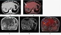 Fig. 3 A 66-year-old man with hepatocellular carcinoma, showing a large hepatic mass with
necrosis, radio-dense foci (post-TARE status), heterogeneous peripheral enhancement,
and adjacent mild FDG uptake anteriorly as can be seen in PET/CT images (A) and (B). Axial T2W images (C) of [18F] FDG PET/MRI scans revealed a large hepatic mass showing heterogeneous FDG
uptake in fused images with two small FDG avid nodular T2 mildly hyperintense lesions
anterolateral to hepatic mass. (D) ADC sequence showing two small lesions showing hypointense signals. (E) PET/MRI showing two small adjacent lesions in segment VIII which was not visible
in PET/CT image. ADC, apparent diffusion coefficient; CT, computed tomography; FDG,
fluoro-D-glucose; MRI, magnetic resonance imaging; PET, positron emission tomography.
Fig. 3 A 66-year-old man with hepatocellular carcinoma, showing a large hepatic mass with
necrosis, radio-dense foci (post-TARE status), heterogeneous peripheral enhancement,
and adjacent mild FDG uptake anteriorly as can be seen in PET/CT images (A) and (B). Axial T2W images (C) of [18F] FDG PET/MRI scans revealed a large hepatic mass showing heterogeneous FDG
uptake in fused images with two small FDG avid nodular T2 mildly hyperintense lesions
anterolateral to hepatic mass. (D) ADC sequence showing two small lesions showing hypointense signals. (E) PET/MRI showing two small adjacent lesions in segment VIII which was not visible
in PET/CT image. ADC, apparent diffusion coefficient; CT, computed tomography; FDG,
fluoro-D-glucose; MRI, magnetic resonance imaging; PET, positron emission tomography.
PET/MRI Shows Better Lesion Delineation Compared with PET/CT
Not only does PET/MRI help reveal additional PET-positive liver lesions but it has
also supported better lesion delineation as compared with PET/CT, which further improves
patient treatment management.
A case of breast carcinoma with liver metastases demonstrated the advantage of PET/MRI
over PET/CT owing to better lesion delineation with MR images. This can be seen in
[Fig. 4], where T2W MR images have demonstrated improved lesion anatomical delineation as
compared with the CT images.
 Fig. 4 Case of breast cancer with liver metastases: Axial CT and simultaneous PET/CT images
are shown in (A) and (B), respectively, and axial T2W MR and simultaneous PET/MRI are shown in (C) and (D), respectively, with improved lesion delineation seen in PET/MR images, highlighted
by blue arrows. PET/MR images show bilobed T2 hyperintense lesion (blue arrow) in
segment VIII of liver with FDG uptake which was minimal and vague on PET/CT images
(A, B), highlighted with an orange arrow. CT, computed tomography; FDG, fluoro-D-glucose;
MRI, magnetic resonance imaging; PET, positron emission tomography; T2W, T2-weighted.
Fig. 4 Case of breast cancer with liver metastases: Axial CT and simultaneous PET/CT images
are shown in (A) and (B), respectively, and axial T2W MR and simultaneous PET/MRI are shown in (C) and (D), respectively, with improved lesion delineation seen in PET/MR images, highlighted
by blue arrows. PET/MR images show bilobed T2 hyperintense lesion (blue arrow) in
segment VIII of liver with FDG uptake which was minimal and vague on PET/CT images
(A, B), highlighted with an orange arrow. CT, computed tomography; FDG, fluoro-D-glucose;
MRI, magnetic resonance imaging; PET, positron emission tomography; T2W, T2-weighted.
PET/MRI Reveals Additional Information over PET/CT
A case of HCC has shown further the benefit of PET/MRI where PET/MRI has revealed
additional information in the form of relation of hepatic mass with the adjacent organs
as compared with PET/CT. As can be seen in [Fig. 5A, B], coronal PET/CT reveals a large hepatic mass predominantly in the right lobe and
segment IV-B with partial exophytic component. Further, coronal PET/MR images as in
[Fig. 5C–E] showed relation of large hepatic mass with the adjacent antrum of stomach, common
hepatic duct (CHD), common bile duct (CBD), gallbladder, and duodenum. This information
was not evident on PET/CT; the additional findings from PET/MRI supported more accurate
patient management.
 Fig. 5 Case of hepatocellular carcinoma abutting adjacent structures: This figure shows
the advantage of PET/MRI in revealing additional information about the hepatic mass
as compared with PET/CT. (A) and (B) show coronal CT and simultaneous PET/CT images, respectively; (C), (D), and (E) show coronal T2W HASTE MR and simultaneous PET/MR images, respectively; (F) shows the histopathology image from true-cut biopsy of FDG avid portion of hepatic
lesion. Coronal PET/CT images, (A) and (B), reveal a large hepatic mass predominantly in the right lobe and segment IV-B with
partial exophytic component. Coronal PET/MR images (C–E) showing relation of large hepatic mass with the adjacent antrum of stomach, CHD,
CBD, gallbladder and duodenum. (F) Histopathology and immunohistochemistry reports revealed neoplastic cells arranged
in nests and sheets with cells showing pleomorphic hyperchromatic nuclei with mitosis,
necrosis, and high Ki-67 index (75–80%). These features are consistent with hepatocellular
carcinoma. CBD, common bile duct; CHD, common hepatic duct; CT, computed tomography;
FDG, fluoro-D-glucose; MRI, magnetic resonance imaging; PET, positron emission tomography.
Fig. 5 Case of hepatocellular carcinoma abutting adjacent structures: This figure shows
the advantage of PET/MRI in revealing additional information about the hepatic mass
as compared with PET/CT. (A) and (B) show coronal CT and simultaneous PET/CT images, respectively; (C), (D), and (E) show coronal T2W HASTE MR and simultaneous PET/MR images, respectively; (F) shows the histopathology image from true-cut biopsy of FDG avid portion of hepatic
lesion. Coronal PET/CT images, (A) and (B), reveal a large hepatic mass predominantly in the right lobe and segment IV-B with
partial exophytic component. Coronal PET/MR images (C–E) showing relation of large hepatic mass with the adjacent antrum of stomach, CHD,
CBD, gallbladder and duodenum. (F) Histopathology and immunohistochemistry reports revealed neoplastic cells arranged
in nests and sheets with cells showing pleomorphic hyperchromatic nuclei with mitosis,
necrosis, and high Ki-67 index (75–80%). These features are consistent with hepatocellular
carcinoma. CBD, common bile duct; CHD, common hepatic duct; CT, computed tomography;
FDG, fluoro-D-glucose; MRI, magnetic resonance imaging; PET, positron emission tomography.
[Fig. 6] also highlights the advantage of PET/MRI in providing additional information about
the hepatic mass compared with PET/CT. PET/MRI reveals a lesion in segment IV of the
liver with mild FDG uptake, attributed to longer PET acquisition time and better spatial
resolution, as seen in [Fig. 6C, D]. Additionally, dynamic contrast-enhanced PET/MRI demonstrates an FDG-avid peripherally
enhancing hepatic lesion and an FDG-avid colonic lesion, which were not visible on
PET/CT ([Fig. 6E]).
 Fig. 6 A 48-year-old man with colonic mass and liver lesions: This figure shows the advantage
of PET/MRI in revealing additional information about the hepatic mass compared with
PET/CT. (A) and (B) show axial CT and simultaneous PET/CT images, respectively; (C) and (D) show axial T2 FSE FS MR and simultaneous PET/MR images, respectively; (E) and (F) show fused contrast-enhanced PET/MRI and venous-phase MR images. PET/MRI shows lesion
in segment IV of the liver with mild FDG uptake due to longer PET acquisition time
and better spatial resolution as can be seen in (C) and (D). Further, DCE PET/MRI showed FDG avid peripherally enhancing hepatic lesion and
FDG avid colonic lesion which was not visible in PET/CT as can be seen in (E). CT, computed tomography; DCE, dynamic contrast-enhanced; FDG, fluoro-D-glucose;
MRI, magnetic resonance imaging; PET, positron emission tomography.
Fig. 6 A 48-year-old man with colonic mass and liver lesions: This figure shows the advantage
of PET/MRI in revealing additional information about the hepatic mass compared with
PET/CT. (A) and (B) show axial CT and simultaneous PET/CT images, respectively; (C) and (D) show axial T2 FSE FS MR and simultaneous PET/MR images, respectively; (E) and (F) show fused contrast-enhanced PET/MRI and venous-phase MR images. PET/MRI shows lesion
in segment IV of the liver with mild FDG uptake due to longer PET acquisition time
and better spatial resolution as can be seen in (C) and (D). Further, DCE PET/MRI showed FDG avid peripherally enhancing hepatic lesion and
FDG avid colonic lesion which was not visible in PET/CT as can be seen in (E). CT, computed tomography; DCE, dynamic contrast-enhanced; FDG, fluoro-D-glucose;
MRI, magnetic resonance imaging; PET, positron emission tomography.
Quantitative Comparison between PET/MRI and PET/CT
In this study, a newly customized liver MR protocol was implemented in conjunction
with quantitative PET to evaluate hepatic lesions using PET/MRI within a concise time
frame of 20 minutes. The utilization of PET/MRI yielded additional information in
87% of cases and identified extra lesions in 73% of patients. PET/MRI exhibited improved
lesion delineation compared with PET/CT, enhancing clinical diagnosis ([Fig. 4]) and facilitating better patient management in 80% of the total studied cases. Notably,
in [Fig. 5C–E], PET/MRI provided crucial supplementary information regarding the relationship between
a large hepatic mass and adjacent structures, such as the gallbladder, duodenum, CHD,
and CBD, which was only partially visualized on PET/CT images ([Fig. 5A, B]). Furthermore, PET/MRI enhanced the identification of supplementary subcentimeter
liver lesions as can be seen in [Figs. 2] and [3], underscoring its superior sensitivity in lesion detection compared with PET/CT.
In the patient cohort, PET/MRI could detect an impressive 40 additional lesions as
opposed to PET/CT. These lesions were as small as 2 mm in long-axis diameter. Additionally,
PET/MRI displayed a higher TLR of the SUV as compared with PET/CT, which had a significant
impact on the final reports and influenced therapeutic decisions. The findings from
PET/MRI notably affected the response assessment category in 5 cases and defined malignant
hepatic lesions on staging/restaging scans in 10 out of 15 cases. The study concludes
that PET/MRI offers enhanced diagnostic capabilities over PET/CT, providing valuable
insights into hepatic lesions and improving patient outcomes.
MR Sequences Enhance Lesion Detectability in PET/MRI
Among the sequences included in the protocol, the T2 FSE FAT SAT sequence demonstrated
superior delineation of small liver lesions. This sequence employs a T2-weighted fast
spin-echo sequence with fat saturation, which effectively suppresses fat signal, thereby
enhancing the visualization and characterization of lesions within the liver. Furthermore,
the late arterial phase contrast imaging showed better delineation of small subcentimeter
liver lesions as compared with early arterial phase contrast imaging.
The study emphasized the significant value of the spatial resolution of MRI and the
complementary data acquired simultaneously by PET/MRI in evaluating lesion viability.
The higher spatial resolution of MRI allows for clearer visualization of lesion characteristics
and provides additional information for precise diagnosis. Additionally, advanced
MR techniques such as DWI and ADC mapping contributed to clinical lesion diagnosis,
further enhancing the diagnostic accuracy of PET/MRI.
Discussion
Accurate identification of hepatic lesions is crucial for optimal therapy and improved
patient outcomes. Hence, it becomes crucial to choose an imaging modality that offers
the highest level of accuracy and precision.[10] Both PET-MR and PET-CT have become valuable tools in hepatic lesion detection, providing
improved diagnostic capabilities. This extensive study thoroughly evaluated the efficacy
of PET/MR and PET/CT in detecting and characterizing hepatic lesions, with a particular
emphasis on 18F-FDG–avid lesions. Although many studies have suggested PET/CT to be
more sensitive than conventional CT in detecting hepatic lesions,[13] due to several limitations, PET/MR scans are increasingly recommended to evaluate
the efficiency and precision of hepatic lesion detection. These scans are suggested
to be done alone or as a delayed scan after the initial PET/CT examination.
PET/MR stands out as a powerful and advanced tool for lesion detection, particularly
in the realm of initial tumor staging. This hybrid imaging modality exhibits a synergistic
effect, combining the metabolic information from PET with the detailed anatomical
and soft tissue characterization provided by MRI.[14] One major benefit of PET/MR compared with PET/CT is the substantial decrease in
radiation exposure, making it a great choice for patients who require multiple imaging
sessions. The combined PET/MR images demonstrate a notable enhancement in sensitivity
and specificity for identifying malignancies in comparison to conducting separate
MRI and PET scans. Accurate tumor staging and treatment planning rely heavily on this
enhanced extent of precision. In addition to tumor staging, PET/MRI shows promising
potential in assessing local lesions, leveraging the innate benefits of MRI. The extensive
anatomical information provided by MRI improves the accuracy of evaluating the extent
of invasion, giving clinicians a thorough understanding of the scope of the lesion.[10]
In the context of hepatic neoplastic lesions, a common application of PET/MRI, complete
and accurate detection becomes paramount, especially in diseases such as colon or
rectal cancer, where therapeutic decisions hinge on factors such as the size and number
of lesions. The amalgamation of a diagnostic multiphase MRI of the liver, which is
widely accepted as the current standard of care, with PET evaluation offers a distinct
and valuable opportunity. This combination not only provides enhanced sensitivity
and specificity but also enables a more thorough evaluation of the likelihood and
viability of distant disease in the liver.[9]
This study demonstrated that PET/MRI, when compared with PET/CT, exhibited superior
performance in terms of lesion delineation and anatomic allocation of PET-positive
findings. It is worth mentioning that the addition of the diagnostic T2 FSE FAT SAT
sequence in PET/MR was critical in achieving better delineation of small liver lesions.
PET/MRI showcased enhanced capabilities in precisely defining the boundaries and anatomical
locations of the lesions over PET/CT. This advantage in anatomic delineation is consistently
reflected in the improved classification of hepatic lesions, emphasizing the clinical
significance of PET/MRI in providing more accurate spatial information for these lesions.
It is worth mentioning that PET/MRI yielded valuable additional insights in 87% of
cases (13 out of 15), enhancing our general comprehension of the disease. In 73% of
patients (11 out of 15), PET/MRI managed to successfully detect additional lesions
that were not identified by PET/CT. This highlights the enhanced sensitivity of PET/MRI
in detecting lesions, which is essential for precise staging and planning of treatment.
The additional lesion findings changed treatment planning for 20% of the patients
where PET/CT only detected one lesion, whereas PET/MRI detected multiple lesions leading
to the change in treatment and patient management. In some patients, detection of
additional lesions on PET/MRI altered treatment plans, including surgical management.
The enhanced lesion delineation observed in 80% of the total studied cases further
emphasizes the superiority of PET/MRI over PET/CT. This improved delineation not only
helps with the precise identification of abnormalities but also has important implications
for clinical diagnosis and subsequent patient care. Particularly noteworthy is the
identification of supplementary subcentimeter liver lesions facilitated by PET/MRI,
emphasizing its capability to detect smaller lesions that might be overlooked by other
imaging modalities.
When it comes to lesion detection, PET/MRI proved to be more effective than PET/CT
by detecting an additional 40 lesions in the patient group. A tiny lesion, measuring
only 2 mm in long-axis diameter, has been effectively detected, illustrating the remarkable
sensitivity of PET/MRI in identifying even the smallest abnormalities. Across 15 patients,
the average long-axis diameter of small lesions measured 3.4 mm, with a standard deviation
of 1.3 mm. This emphasizes the accuracy of PET/MRI in analyzing lesions of different
sizes. In addition, the data we acquired revealed that PET/MRI had a higher TLR compared
with PET/CT. This metric is critical in assessing the metabolic activity of lesions,
providing valuable insights into their biological characteristics.
The reconstruction of PET datasets using a 3D OSEM and HYPER Deep Progressive Reconstruction
Algorithm, along with the automatic generation of a four-compartment-model attenuation
map (μ-map) based on a water-fat-imaging sequence with breath gating for PET/MRI,
contributed to the accuracy and reliability of the results obtained. This robust methodology
ensures precise attenuation correction, enhancing the overall diagnostic capability
of PET/MRI in hepatic lesion evaluation. In conclusion, the comprehensive results
of this study affirm that PET/MRI is a superior imaging modality, offering enhanced
lesion detection, delineation, and characterization compared with PET/CT in the evaluation
of hepatic lesions.
A few limitations need to be acknowledged in this study. First, the use of FDG as
a tracer is not tumor-specific, necessitating an understanding of physiological variations
in FDG uptake and potential pitfalls and artifacts. Nonspecific FDG uptake can lead
to false positives or negatives, affecting the accuracy of lesion characterization.
Second, the study primarily consists of a single-center case series, limiting the
generalizability of findings to broader populations.[15] Multicenter studies could provide more robust evidence of the general applicability
of PET/MRI in hepatic lesion evaluation. Finally, in the detection of 18F-FDG–negative
lesions, the performance of the CT versus MRI components may differ significantly,
potentially influencing the overall sensitivity and specificity of the imaging modality.
Awareness of these limitations is crucial for the judicious interpretation of results
and underscores the need for further research to address these challenges.
Conclusion
In conclusion, our study provides compelling evidence supporting the significant advantages
of PET/MRI over PET/CT in the detection and characterization of hepatic lesions. One
notable advantage is the substantial reduction in ionizing radiation exposure by 80%,
reinforcing PET/MRI as a safer alternative for patients requiring repeated imaging
sessions. Additionally, the study revealed higher detection rates and more precise
characterization of small malignant liver lesions with the PET/MRI combination, emphasizing
its superior clinical utility.
The incorporation of the T2 FSE FAT SAT MR sequence in PET/MRI demonstrated a higher
rate of concordant findings compared with PET/CT. This heightened concordance, particularly
in the context of smaller lesions, not only enhances diagnostic certainty but also
has direct implications for patient management. PET/MRI emerges as a powerful imaging
tool, offering a more comprehensive and accurate assessment that directly impacts
patient diagnosis, staging, and subsequent therapeutic strategies.[9]
These findings underscore the potential of PET/MRI to serve as a transformative imaging
technology in oncologic imaging, providing clinicians with enhanced diagnostic confidence
and valuable insights for individualized patient care. As we move forward, the utilization
of PET/MRI, with its notable reduction in radiation exposure and improved diagnostic
performance, has the potential to reshape the landscape of hepatic lesion evaluation,
influencing clinical decision-making and ultimately improving patient outcomes. Further
research and broader multicenter studies will be crucial to solidify these findings
and establish PET/MRI as a standard in hepatic lesion detection and characterization.


