

Subscribe to RSS
DOI: 10.1055/s-2007-995810
© Georg Thieme Verlag KG Stuttgart · New York
Cervical esophageal capillary hemangioma removed by combined and sequential endoscopic ligation and snare polypectomy
H. H. Wang, MD
Peking University First Hospital
8 Xishiku street
Beijing, 100034
China
Fax: +86-10-66518105
Email: wanghuahong@medmail.com.cn
Publication History
Publication Date:
30 July 2008 (online)

Esophageal capillary hemangioma is a rare benign esophageal tumor, and is usually asymptomatic [1]. More and more patients with esophageal hemangioma are being treated endoscopically [2] [3] by combined techniques in order to prevent massive bleeding, although the advantages are still controversial.
A 68-year-old Chinese male, complaining of intermittent pharyngeal obstruction, was found to have a round, smooth protruding lesion with almost normal esophageal mucosa (19 cm from the incisors) by routine esophagogastroduodenoscopy (Olympus XQ240, Japan) ([Fig. 1]). Contrast-enhanced computed tomography (CT) showed an irregular intramural mass lesion in the cervical esophagus, 0.7 × 0.6 cm in size. There was marked enhancement following intravenous contrast (CT value = 184.61 Hu), and high density on delayed scan (CT value = 98.9 Hu) ([Fig. 2]). In order to prevent massive bleeding, we decided to perform combined and sequential endoscopic ligation and snare polypectomy, in order to remove the lesion endoscopically. The patient signed the informed consent form.

Fig. 1 Endoscopy revealed a round, smooth protruding lesion covered by an almost normal esophageal mucosa at 19 cm apart from the incisor.
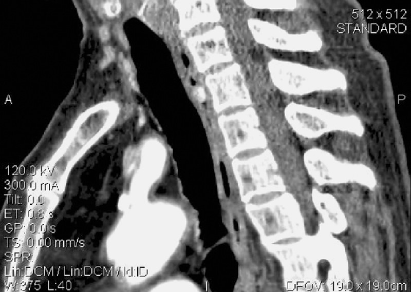
Fig. 2 Contrast-enhanced computed tomography (CT) showed an intramural mass lesion in the cervical esophagus, 0.7 × 0.6 cm in size, with marked enhancement following intravenous contrast (CT value = 184.61 Hu) and high density on delayed scan (CT value = 98.9 Hu).
In brief, the tumor was aspirated into a hood cap attached to the top of the endoscope and ligated with an O-ring in a manner similar to that used for endoscopic variceal ligation (Saeed ligator, Wilson-Cook Medical GI Endoscopy, USA) ([Fig. 3 ] a). One week later, the tumor was not so fragile and was removed by snare polypectomy ([Fig. 3 ] b). No bleeding occurred and the base of the cutting was clear. After removal of the lesion, the symptoms disappeared. Pathology showed lobulated capillary hemangioma with ulceration ([Fig. 4]) and strong positive CD34 staining ([Fig. 5]). No remarkable thrombosis and necrosis were found pathologically. No recurrence was evident after 2 months. We did notice that prior ligation can facilitate the procedure of snare polypectomy, which is the advantage of the combined technique over the simple snare polypectomy.


Fig. 3 a The lesion appearance 1 week after ligation and before polypectomy; b The mucosal appearance after snare polypectomy.

Fig. 4 The pathological examination showed lobulated capillary hemangioma (hematoxylin & eosin, × 200).

Fig. 5 The immunohistochemical test showed strong positive staining for CD34 (× 200).
In conclusion, combined and sequential ligation and snare polypectomy is a good option for the endoscopic removal of esophageal protruding hemangioma. But the efficacy and advantages of prior ligation still need more investigation.
Endoscopy_UCTN_Code_TTT_1AO_2AG
References
- 1 Cantero D, Yoshida T, Ito T. et al . Esophageal hemangioma: endoscopic diagnosis and treatment. Endoscopy. 1994; 26 250-253
- 2 Tominaga K, Arakawa T, Ando K. et al . Oesophageal cavernous haemangioma diagnosed histologically, not by endoscopic procedures. J Gastroenterol Hepatol. 2000; 15 215-219
- 3 Sogabe M, Taniki T, Fukui Y. et al . A patient with esophageal hemangioma treated by endoscopic mucosal resection: a case report and review of the literature. J Med Invest. 2006; 53 177-182
H. H. Wang, MD
Peking University First Hospital
8 Xishiku street
Beijing, 100034
China
Fax: +86-10-66518105
Email: wanghuahong@medmail.com.cn
Fig. 1 Endoscopy revealed a round, smooth protruding lesion covered by an almost normal esophageal mucosa at 19 cm apart from the incisor.
Fig. 2 Contrast-enhanced computed tomography (CT) showed an intramural mass lesion in the cervical esophagus, 0.7 × 0.6 cm in size, with marked enhancement following intravenous contrast (CT value = 184.61 Hu) and high density on delayed scan (CT value = 98.9 Hu).
Fig. 3 a The lesion appearance 1 week after ligation and before polypectomy; b The mucosal appearance after snare polypectomy.
Fig. 4 The pathological examination showed lobulated capillary hemangioma (hematoxylin & eosin, × 200).
Fig. 5 The immunohistochemical test showed strong positive staining for CD34 (× 200).