
Subscribe to RSS
Please copy the URL and add it into your RSS Feed Reader.
https://www.thieme-connect.de/rss/thieme/en/10.1055-s-00000041.xml
Neuropediatrics 1992; 23(1): 24-27
DOI: 10.1055/s-2008-1071306
DOI: 10.1055/s-2008-1071306
Original article
© Georg Thieme Verlag KG Stuttgart · New York
Computed Cranial Tomography, Magnetic Resonance Imaging and Single Photon Emission Computed Tomography in Hemorrhagic Shock and Encephalopathy Syndrome: A Report of Three Cases
Further Information
Publication History
Publication Date:
19 March 2008 (online)
Abstract
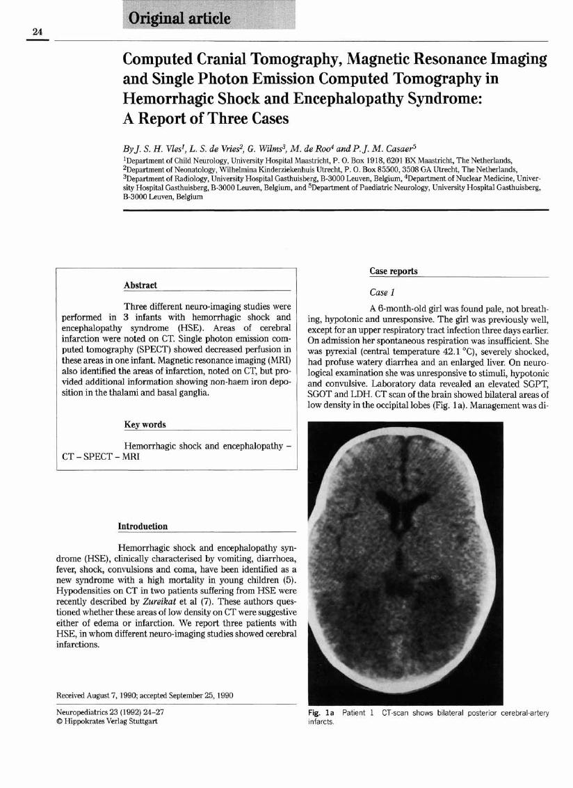
Three different neuro-imaging studies were performed in 3 infants with hemorrhagic shock and encephalopathy syndrome (HSE). Areas of cerebral infarction were noted on CT. Single photon emission computed tomography (SPECT) showed decreased perfusion in these areas in one infant. Magnetic resonance imaging (MRI) also identified the areas of infarction, noted on CT, but provided additional information showing non-haem iron deposition in the thalami and basal ganglia.
Key words
Hemorrhagic shock and encephalopathy - CT - SPECT - MRI