

Subscribe to RSS
DOI: 10.1055/a-1930-9714
Subclavian Effort and Upper Limb Thrombosis – a Lesson Learned
Effort-Thrombose der Vena subclavia und Thrombose der oberen Gliedmaßen – eine LektionAuthors

Abstract
Subclavian vein effort and upper limb thrombosis, known as the Paget-Schroetter syndrome (PSS), accounts for 30–40 % of spontaneous upper extremity deep vein thromboses (UEDVTs) and 10–20 % of all upper limb deep vein thromboses (DVTs). As complication of PSS include post-thrombotic syndrome and pulmonary embolism, early recognition and prompt initiation of anticoagulant treatment is crucial in the course of its treatment. PSS is associated with single or repeated physical activity of the upper limb, combined with obstruction of venous outflow resulting from anatomical alterations. A correct diagnosis, based on a range of imaging methods, and prompt initiation of local thrombolytic therapy, surgical decompression of the thoracic outlet (when necessary), and immediate initiation of anticoagulant treatment, aim to effectively restore the patient life quality, preventing post-thrombotic syndrome and recurrent thrombosis.
Zusammenfassung
Die Effort-Thrombose der Vena subclavia und die Thrombose der oberen Gliedmaßen werden als Paget-Schroetter-Syndrom (PSS) bezeichnet. Sie machen 30–40% der spontanen tiefen Venenthrombosen der oberen Extremitäten und 10–20% aller tiefen Venenthrombosen der oberen Extremitäten aus. Zu den Komplikationen des PSS werden das postthrombotische Syndrom und die Lungenembolie gezählt. Daher sind das frühzeitige Erkennen und die unverzügliche Einleitung einer gerinnungshemmenden Behandlung für den Verlauf von entscheidender Bedeutung. Das PSS tritt bei einmaliger oder wiederholter körperlicher Betätigung der oberen Gliedmaßen in Verbindung mit einer Obstruktion des venösen Abflusses infolge anatomischer Veränderungen auf. Eine korrekte Diagnose auf der Grundlage verschiedener bildgebender Verfahren und die unverzügliche Einleitung einer lokalen Thrombolysetherapie – einer chirurgischen Dekompression des Thoraxauslasses (falls erforderlich) und die sofortige Einleitung einer gerinnungshemmenden Behandlung – sollen die Lebensqualität des Patienten wirksam wiederherstellen und ein postthrombotisches Syndrom sowie eine erneute Thrombose verhindern.
Schlüsselwörter
Tiefe Venenthrombose der oberen Extremitäten - Thoracic-Outlet-Syndrom - Thrombose der Vena subclavia - Paget-Schroetter-Syndrom - Thrombolyse - Resektion der ersten RippeKeywords
upper extremity deep vein thrombosis - thoracic outlet syndrome - subclavian vein thrombosis - Paget-Schroetter syndrome - thrombolysis - first rib resectionIntroduction
Primary “spontaneous” upper extremity deep vein thrombosis (DVT) is a rare disease comprising thrombosis of the deep veins draining the upper extremity. It results from anatomical abnormalities of the thoracic outlet, which cause axillo-subclavian compression and subsequent thrombosis. While this condition is commonly known as the venous thoracic outlet syndrome (TOS), it is also referred to as the Paget-Schroetter syndrome, as well as the "effort" thrombosis [1].
As the name of the syndrome indicates, the venous thoracic outlet syndrome (vTOS) is a severe stenosis or thrombosis of the subclavian-axillary vein, resulting from chronic extrinsic mechanical compression. First described by Paget (1875) and Schroetter (1884), Hughes named this condition the “Paget-Schroetter syndrome” (PSS) in 1949, in a comprehensive literature review regarding thrombosis of the subclavian and axillary veins [2]. The most recent name for venous TOS (vTOS) is effort thrombosis, taking into account its relatively common presence among young healthy individuals engaging in activities that require repetitive arm and shoulder motion [3]. Different subtypes of venous TOS were presented in [Fig. 1].

While PSS is more frequently reported in young, healthy, athletic males (mean age 15–30 years), the number of women burdened by this condition is also increasing. Two aspects often characterize PSS, namely young age of patients and its presence in the dominant extremity, maximum effort should be taken to diagnose and treat it as soon as possible. Moreover, a thorough understanding the thoracic outlet is crucial to efficiently treat patients, as the subclavian vein is located in a compartment separated from the subclavian artery and brachial plexus by the anterior scalene muscle. Hence, compression of the subclavian vein by the musculoskeletal components of the thoracic outlet or costoclavicular space may result in acute thrombosis.
Pathogenesis
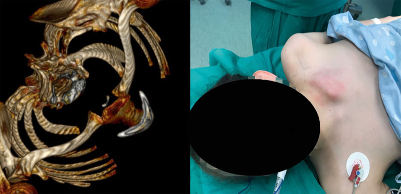
Subclavian vessels and the shoulder plexus pass through an anatomically narrow space formed by the anterior and middle scalene muscles, the 1st rib, the clavicle and the attachment of the pectoralis minor muscle. Out of all the vessels, the subclavian vein is most likely to be compressed. Alterations of the structures forming this space (congenital or acquired), leading to its restriction, result in compression of the subclavian artery and/or subclavian vein and the brachial plexus. This occurrence can particularly often be observed in people who perform repeated actions that require inversion or elevation of the upper limb (wall painters, electricians, conductors, hairdressers). Moreover, congenital anomalies playing a role in PSS include cervical ribs, supernumerary muscles, abnormal tendon insertions, or abnormal muscular or tendinous bands [4]. As an example, in [Fig. 2], a congenital anomaly in the form of a fused 1st and 2nd rib of a 14-year-old girl can be observed. In turn, acquired abnormalities resulting in PSS include bone overgrowth due to fracture (e.g., of the clavicle or first rib), or hypertrophy of anterior scalene subclavius muscles, often related to repetitive limb lifting [5].
Subclavian vein thrombosis develops as a consequence of repetitive microtrauma resulting from compression characteristic for the thoracic outlet syndrome (TOS).
Repeated trauma during limb movement leads to thickening and fibrosis within the venous vessel walls. This results in changes to the tunica intima, the surface of which becomes thrombogenic. In the case of the venous system, stenosis most commonly occurs at the junction of the brachiocephalic and subclavian veins. Hence, compression-associated blood flow impairment and endothelial changes resulting from repeated trauma can result in thrombosis of the subclavian and/or axillary vein.
Epidemiology
It is estimated that venous thromboembolic diseases annually manifest in approximately 200–300 in 100,000 persons. One-third of these patients presents a pulmonary embolism (PE), while two-thirds are diagnosed with deep vein thrombosis (DVT) [6].
Depending on the etiology, upper limb DVT can be classified as either primary or secondary. Furthermore, PSS accounts for 30–40% of spontaneous UEDVT cases, and for 10–20 % of all upper extremity deep venous thromboses [7].
Primary thrombosis of the subclavian vein often follows heavy physical effort and affects people at a younger age, particularly men. In contrast, secondary thrombosis occurs as a result of trauma, neoplastic processes, the presence of catheters in the venous lumen and the use of oral contraceptives. Upper thoracic outlet syndrome refers to compression of the neurovascular structures extending from the thorax towards the upper limb. TOS represents the sum of clinical symptoms, regardless of the cause of the compression of the nerves and vessels of the upper limb, starting from the level of the neck down to the axillary fossa. TOS occurs in approximately 10 in 100,000 people [6]. Furthermore, venous TOS affects 3–5% of patients and is diagnosed more often in men than in women, occurs mostly in young people, and is usually localised in the dominant arm [8].
Risk factors for primary subclavian vein thrombosis include repetitive movements of the upper limb in abduction, often combined with high physical effort. Professional athletes account for approximately 40% of patients undergoing resection of the first rib or anterior scalene muscle due to the presence of neurovascular compression [9] [10]. Other important predisposing factors for UEDVT include indwelling devices (e.g., central venous catheters, ports, and pacemakers), occult or overt malignancies, and other thrombophilic states [7].
Symptoms
In the case of primary subclavian vein thrombosis, symptoms appear early and develop relatively rapidly. In turn, the development and progression of symptoms is slightly slower in the case of secondary thrombosis. Affected patients report a burning pain in the axillary fossa and the medial surface of the upper limb. Swelling of the dorsal surface of the hand, fingers and forearm can also be observed, sometimes affecting the entire upper limb. The skin becomes tensed and takes on a bluish tinge as a result of blood stasis. At a later stage, oedema develops on the anterior surface of the shoulder and chest. The above changes may result in a limitation of limb movement, especially at the shoulder joint. Rarer symptoms include fever, tachycardia, and chills. On examination, the subclavian vein is painful when compressed, and markedly thickened.
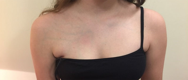
Primary UEDVT is characterized by compression of the axillary or subclavian vein, resulting in blood flow obstruction. Its most important risk factors include specific types of intense exercise or/and anatomical abnormalities, including enlargement of the costoclavicular ligament, located between the clavicle and the first rib. Due to physical activity and subsequent hypertrophy of the surrounding muscles, the vein can be significantly narrowed, and thrombosis can develop. Patients with primary UEDVT exhibit a higher prevalence of antiphospholipid antibody, factor V Leiden, and prothrombin gene mutations compared to the general population [11]. Patients often present oedema, pain, upper limb discoloration, paraesthesia, weakness, or cyanosis. [Fig. 3] presents a 15-year-old girl with right subclavian vein thrombosis, manifesting in oedema and thickening of right-side venous vessels. In turn, PSS epidemiological data and risk factors were presented in [Table 1]. Primary UEDVT is a rare condition, with estimated annual incidence between one and two in 100,000 patients, with an equal distribution between the sexes. In secondary UEDVT, blood clots are most frequently located in the subclavian vein, but are also found in the jugular, axillary, brachial, or brachiocephalic veins. Rarely, more distal veins, such as the radial vein, are involved. Due to the strong association of secondary UEDVT with cancer and CVC, the mean age at presentation is around 60 years, substantially older than in primary UEDVT [11].
Diagnosis
A diagnosis of upper extremity venous outflow obstruction (i.e., deep vein thrombosis or venous stenosis) may be suspected based on the clinical presentation, but should be confirmed using imaging, typically ultrasound (B-mode ultrasound, color Doppler ultrasound, and duplex ultrasound).
Ultrasonography is considered the examination of choice in the diagnosis of UEDVT. A systematic review, based on 17 studies, concluded that compression ultrasonography is an acceptable alternative to standard contrast venography. The summary estimates of the compression sensitivity, Doppler ultrasound, and Doppler ultrasound with compression were 97, 84, and 81 percent, respectively. In turn, specificities of the above-mentioned diagnostic methods were 96, 94, and 93 percent, respectively [12]. Importantly, the examination can be performed dynamically in functional positions of the limb, which increases its diagnostic value. Furthermore, blood flow characteristics can also be determined during the examination, with significant acceleration of flow observed indicating stenosis and compression, while no flow suggests complete closure of the vessel. However, the accuracy of the examination depends on the anatomical conditions of the patient and the experience of the examiner. Disadvantages of ultrasound include technician participation dependence, and the possibility of difficulties in identifying nonocclusive mural thrombi, as well as thrombi in the proximal subclavian or innominate veins, due to acoustic shadowing of the overlying clavicle and sternum. Considering these concerns, imaging studies with the administration of a contrast agent are performed to obtain an unambiguous assessment of thrombotic changes in the veins of the upper limb. This is particularly important in the assessment of “old” venous thrombosis, lasting more than six weeks. CT angio is also a valuable addition to non-invasive ultrasound, allowing for the assessment of compression sites of the proximal subclavian vein. Performing transverse scans can also provide information about the tissue structure (e.g., tumors, lymph nodes, thrombi, vessel walls).
Angio-MR is also a recommended examination in the assessment of venous compression syndromes caused by muscular-fibrous structures. In our center, we also perform it as a follow-up of first rib removal, to assess the flow in the venous system and the radicality of the surgery. [Fig. 4] shows follow-up NMR (nuclear magnetic resonance) of first left rib excision surgery, with normal flow through the left subclavian vein. Classical phlebography is the oldest and most invasive examination method, due to the need of venous catheter insertion and contrast administration. As a diagnostic test, it is used in doubtful cases of compression and planned venous reconstructions. However, it still remains the “gold standard” for venous pathology evaluation and plays a particularly important role in the intraoperative assessment of blood flow through the subclavian vein, both before and after reconstruction or decompression surgery [6].

Although catheter-based (digital subtraction) venography allows for the best visualization of abnormal venous anatomy and is the standard with which other methods are compared, it is generally not necessary for diagnosis of upper extremity deep vein thrombosis or compression. Because of its invasive nature, catheter-based venography is generally reserved for situations in which high clinical suspicion of a primary cause of venous outlet obstruction is present, and other diagnostic methods remain inconclusive [1]. A complex schematic of UEDVT diagnostic flow can be found in a 2020 publication of Bosch et al. [11]. In this work, UEDVT was classified based on the criteria proposed by Constans et al [13].
Treatment
The Department of Vascular Surgery in Poznan has been performing 1st rib removal surgery for over 30 years [6]. According to the authors, the basic principles of UEDVT treatment include rapid restoration of normal blood flow using anticoagulant prophylaxis, inhibition of the growth of thrombus and the surrounding inflammatory process, and restoration of the original limb function. Prompt removal, and to the greatest extent possible, of thrombotic material can preserve valve function and reduce venous hypertension [14]. Classical anticoagulant DVT treatment is often characterized by delayed effects and unpredictable outcomes, with conservative treatment based on UFH, LMWH or DOAC anticoagulants. On the other hand, when diagnosing primary, post-exertional thrombosis in the course of VTOS in a young patient, in addition to restoring patency of the subclavian vein, we always aim to eliminate the primary cause of venous thrombosis. Consideration should be given to the fact that the patients treated are often of productive age, with the expectation of full limb function that allows them to continue their normally lifestyle. The treatment principle adopted in the Poznan center is that the most effective and appropriate procedure to achieve the above goal is the initial administration of LMWH with simultaneous insertion of a catheter into the subclavian vein to perform phlebography. If thrombosis is confirmed, local administration of catheter-directed thrombolysis (CDT) is initiated. Two primary thrombolytic drugs, urokinase and rtPA, are used nowadays. In our center, rtPA is administered by local infusion at 1 mg/h, with continuous monitoring of fibrinogen levels, maintained at 200 mg/dL, aPTT and thrombocytes. Maximum treatment time is 72 hours, and its indications include severe symptoms, thrombus occupying most of the subclavian and axillary veins, symptoms lasting for 14 days or more, good functional status, life expectancy of at least 1 year, and low risk of hemorrhage [15]. We also agree with the additional condition posed by the NCCN guidelines, recommending CDT in appropriate candidates, based on institutional expertise [16].
In rare cases, we use more aggressive treatment in the form of percutaneous pharmacomechanical thrombectomy (PMT). This method combines local administration of a fibrinolytic agents and mechanical thrombus removal. The advantages of this procedure over CDT include instant bypass reduction of embolic material and the possibility of returning blood flow immediately after the procedure.
At the Poznan center, the necessary condition for UEDVT treatment, apart from restoring blood flow as quickly as possible, is to remove the cause of subclavian vein compression by excision of the first rib with the adjacent muscular-fibrous structures. This procedure is most often performed during the same hospital stay. In studies on interventional PSS treatment, many authors consider performing this procedure at a deferred date, e.g., within 4–6 weeks after fibrinolytic treatment [10].
More than 30 years of experience based on hundreds of operated patients allows us to recommend the PSS treatment principle in the form of rapid restoration of venous blood flow by surgical removal of the causative factor and endovascular lesions of the subclavian vein if they significantly restrict flow after excision of the first rib. An analysis by Vazquez et al. based on the outcomes of 1271 patients with UEDVT, was published in 2017, indicating excision of the first rib as necessary in all patients [17]. The primary evaluation criterion was the occurrence of PTS, while secondary criteria comprised recurrence of DVT, PE, death, hemorrhage and the need for reoperation with a possibility of endovascular procedure. In their final conclusions, the authors stated that anticoagulant treatment alone does not protect the patient from the occurrence of PTS. This also confirms our observations that surgical treatment is necessary to improve final outcomes, which translates into patients' quality of life, less frequent recurrence of DVT and lower risk of post-thrombotic syndrome [17].
There are three main surgical access points in subclavian vein decompression. The first one comprises 1st rib excision surgery with simultaneous, partial removal of the anterior scalene muscle through the axillary fossa. The advantage of this method is the small cosmetic postoperative scar and the possibility of creating a wide space (after rib excision) for the passage of vascular and nerve structures. Its disadvantages include small surgical field and the inability to directly reconstruct the vein damaged by compression. The authors mostly use this access in adolescent patients.
The second method, subclavian access, is characterized by a better surgical field, allowing for wide exposure of the subclavian vein after excision of the subclavian muscle, the costoclavicular ligament and removal of the anterior part of the 1st rib. This access method facilitates the reconstruction of the vessel using the patient's saphenous vein, as well as the insertion of a venous or biological patch to widen the lumen of the vessel. Both methods presented yield similar final results, with 75–80 % of VTOS patients remaining asymptomatic with an obstructed subclavian vein [18].
A third option is a simultaneous incision above and below the clavicle. The good control of the anatomy obtained by this method increases the extent and radicality of the resected bone-muscle-tendon structures compressing the vein. Oral anticoagulants are recommended for a minimum of three months following such surgery. With such aggressive approach, success rates for re-establishing subclavian vein patency are nearly 100%, provided that thrombolysis is performed within two weeks of symptom onset [19] [20].
It is very important not to perform stenting of the subclavian vein in patients without prior resection of the first rib. Stenting of the vein passing through the non-decompressed costoclavicular junction is often complicated by stent fracture, deformation, and high re-thrombosis rate. [Fig. 5] shows a damaged stent implanted in a patient without prior resection of the first rib.

If the patient does not agree to surgery, we recommend, in accordance with the International Society on Thrombosis and Haemostasias (ISTH), administration of long-term anticoagulant treatment, especially for primary UEDVT with a tight thoracic outlet that has not been surgically corrected [21].
In conclusion, an aggressive approach involving a combination of thrombolysis and thoracic outlet decompression with or without venoplasty (percutaneous, open) appears to improve long-term outcomes (recurrent thrombosis, post-thrombotic syndrome) and improve quality of life in patients with primary deep vein thrombosis of the upper limbs, especially those with acute, moderate or severe symptoms.
Conflict of Interest
The authors declare that they have no conflict of interest.
-
References
- 1 Illig KA, Doyle AJ. A comprehensive review of Paget-Schroetter syndrome. J Vasc Surg 2010; 51: 1538-1547
- 2 Hughes ES. Venous obstruction in the upper extremity; Paget-Schroetter’s syndrome; a review of 320 cases. Surg Gynecol Obstet 1949; 88: 89-127
- 3 Kommareddy A, Zaroukian MH, Hassouna HI. Upper extremity deep venous thrombosis. Semin Thromb Hemost 2002; 28: 89-99
- 4 Fisher JB, Granson MA. Congenital venous web causing subclavian vein obstruction: a case report. J Vasc Surg 1989; 10: 460
- 5 Yamashita Y, Morimoto T, Amano H. et al. Deep vein thrombosis in upper extremities: Clinical characteristics, management strategies and long-term outcomes from the COMMAND VTE Registry. Thromb Res 2019; 177: 1-9
- 6 Pukacki P, Juszkat R, Błaszyk M. et al. Upper extremity deep vein thrombosis: pathogenesis and treatment. Acta Angiol 2019; 25: 115-119
- 7 Alla VM, Natarajan N, Kaushik M. et al. Paget-schroetter syndrome: review of pathogenesis and treatment of effort thrombosis. West J Emerg Med 2010; 11: 358-362
- 8 Hussain MA, Aljabri B, Al-Omran M. Vascular Thoracic Outlet Syndrome. Semin Thorac Cardiovasc Surg 2016; 28: 151-157
- 9 Machleder H. The anterior scalene muscle in thoracic outlet compression syndrome. Archives of Surgery 1986; 121: 1141
- 10 Chang KZ, Likes K, Demos J. et al. Routine venography follow- ing transaxillary first rib resection and scalenectomy (FRRS) for chronic subclavian vein thrombosis ensures excellent outcomes and vein patency. Vasc Endovascular Surg 2012; 46: 15-20
- 11 Bosch FTM, Nisio MD, Büller HR. et al. Diagnostic and Therapeutic Management of Upper Extremity Deep Vein Thrombosis. J Clin Med 2020; 9: 2069
- 12 Di Nisio M, Van Sluis GL, Bossuyt PM. et al. Accuracy of diagnostic tests for clinically suspected upper extremity deep vein thrombosis: a systematic review. J Thromb Haemost 2010; 8: 6840
- 13 Constans J, Salmi L, Sevestre-Pietri MA. et al. A clinical prediction score for upper extremity deep venous thrombosis. Thromb Haemost 2008; 99: 202-207
- 14 Vedantham S. Valvular dysfunction and venous obstruction in the postthrombotic syndrome. Thromb Res 2009; 123: S62-S65
- 15 Kearon C, Akl EA, Comerota AJ. et al. Antithrombotic therapy for VTE disease: Antithrombotic Therapy and Prevention of Thrombosis, 9th ed: American College of Chest Physicians Evidence-Based Clinical Practice Guidelines. Chest 2012; 141 (Suppl. 02) e419S-494S
- 16 Wagman LD, Baird MF, Bennett CL. et al. Venous thromboembolic disease. NCCN. Clinical practice guidelines in oncology. J Natl Compr Canc Netw 2008; 6: 716-753
- 17 Vazquez FJ, Paulin P, Poodts D. et al. Preferred management of primary deep arm vein thrombosis. Eur J Vasc Endovasc Surg 2017; 53: 744-751
- 18 Vemuri C, Salehi P, Benarroch-Gampel J. et al. Diagnosis and treatment of effort-induced thrombosis of the axillary subcla- vian vein due to venous thoracic outlet syndrome. J Vasc Surg Venous Lymphat Disord 2016; 4: 485-500
- 19 Molina JE, Hunter DW, Dietz CA. Paget-Schroetter syndrome treated with thrombolytics and immediate surgery. J Vasc Surg 2007; 45: 328
- 20 Schneider DB, Dimuzio PJ, Martin ND. et al. Combination treatment of venous thoracic outlet syndrome: open surgical decompression and intraoperative angioplasty. J Vasc Surg 2004; 40: 599
- 21 Grant JD, Stevens SM, Woller SC. et al. Diagnosis and management of upper extremity deep-vein thrombosis in adults. Thromb Haemost 2012; 108: 1097-1108
Korrespondenzadresse
Publication History
Article published online:
29 November 2022
© 2022. Thieme. All rights reserved.
Georg Thieme Verlag KG
Rüdigerstraße 14, 70469 Stuttgart, Germany
-
References
- 1 Illig KA, Doyle AJ. A comprehensive review of Paget-Schroetter syndrome. J Vasc Surg 2010; 51: 1538-1547
- 2 Hughes ES. Venous obstruction in the upper extremity; Paget-Schroetter’s syndrome; a review of 320 cases. Surg Gynecol Obstet 1949; 88: 89-127
- 3 Kommareddy A, Zaroukian MH, Hassouna HI. Upper extremity deep venous thrombosis. Semin Thromb Hemost 2002; 28: 89-99
- 4 Fisher JB, Granson MA. Congenital venous web causing subclavian vein obstruction: a case report. J Vasc Surg 1989; 10: 460
- 5 Yamashita Y, Morimoto T, Amano H. et al. Deep vein thrombosis in upper extremities: Clinical characteristics, management strategies and long-term outcomes from the COMMAND VTE Registry. Thromb Res 2019; 177: 1-9
- 6 Pukacki P, Juszkat R, Błaszyk M. et al. Upper extremity deep vein thrombosis: pathogenesis and treatment. Acta Angiol 2019; 25: 115-119
- 7 Alla VM, Natarajan N, Kaushik M. et al. Paget-schroetter syndrome: review of pathogenesis and treatment of effort thrombosis. West J Emerg Med 2010; 11: 358-362
- 8 Hussain MA, Aljabri B, Al-Omran M. Vascular Thoracic Outlet Syndrome. Semin Thorac Cardiovasc Surg 2016; 28: 151-157
- 9 Machleder H. The anterior scalene muscle in thoracic outlet compression syndrome. Archives of Surgery 1986; 121: 1141
- 10 Chang KZ, Likes K, Demos J. et al. Routine venography follow- ing transaxillary first rib resection and scalenectomy (FRRS) for chronic subclavian vein thrombosis ensures excellent outcomes and vein patency. Vasc Endovascular Surg 2012; 46: 15-20
- 11 Bosch FTM, Nisio MD, Büller HR. et al. Diagnostic and Therapeutic Management of Upper Extremity Deep Vein Thrombosis. J Clin Med 2020; 9: 2069
- 12 Di Nisio M, Van Sluis GL, Bossuyt PM. et al. Accuracy of diagnostic tests for clinically suspected upper extremity deep vein thrombosis: a systematic review. J Thromb Haemost 2010; 8: 6840
- 13 Constans J, Salmi L, Sevestre-Pietri MA. et al. A clinical prediction score for upper extremity deep venous thrombosis. Thromb Haemost 2008; 99: 202-207
- 14 Vedantham S. Valvular dysfunction and venous obstruction in the postthrombotic syndrome. Thromb Res 2009; 123: S62-S65
- 15 Kearon C, Akl EA, Comerota AJ. et al. Antithrombotic therapy for VTE disease: Antithrombotic Therapy and Prevention of Thrombosis, 9th ed: American College of Chest Physicians Evidence-Based Clinical Practice Guidelines. Chest 2012; 141 (Suppl. 02) e419S-494S
- 16 Wagman LD, Baird MF, Bennett CL. et al. Venous thromboembolic disease. NCCN. Clinical practice guidelines in oncology. J Natl Compr Canc Netw 2008; 6: 716-753
- 17 Vazquez FJ, Paulin P, Poodts D. et al. Preferred management of primary deep arm vein thrombosis. Eur J Vasc Endovasc Surg 2017; 53: 744-751
- 18 Vemuri C, Salehi P, Benarroch-Gampel J. et al. Diagnosis and treatment of effort-induced thrombosis of the axillary subcla- vian vein due to venous thoracic outlet syndrome. J Vasc Surg Venous Lymphat Disord 2016; 4: 485-500
- 19 Molina JE, Hunter DW, Dietz CA. Paget-Schroetter syndrome treated with thrombolytics and immediate surgery. J Vasc Surg 2007; 45: 328
- 20 Schneider DB, Dimuzio PJ, Martin ND. et al. Combination treatment of venous thoracic outlet syndrome: open surgical decompression and intraoperative angioplasty. J Vasc Surg 2004; 40: 599
- 21 Grant JD, Stevens SM, Woller SC. et al. Diagnosis and management of upper extremity deep-vein thrombosis in adults. Thromb Haemost 2012; 108: 1097-1108