

Subscribe to RSS
DOI: 10.1055/s-0037-1620243
Endoscopic Transsphenoidal Resection of Craniopharyngioma
Authors
Address for correspondence
Publication History
16 October 2017
14 December 2017
Publication Date:
16 January 2018 (online)

Abstract
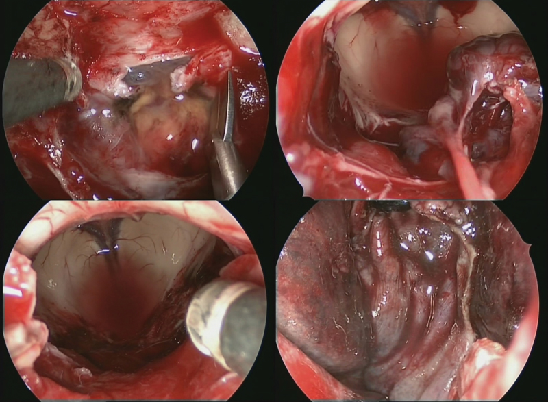
Objectives To demonstrate, step-by-step, the technique and efficacy of endoscopic transsphenoidal approach in resection of a suprasellar craniopharyngioma.
Design The video shows a step-by-step approach to the resection, covering the exposure, access, resection, and confirmation of resection and reconstruction.
Setting The surgery was performed in the University of Malaya Medical Centre, a tertiary referral center in the capital of Malaysia.
Participants Surgery was performed jointly by Professor Prepageran from the department of otorhinolaryngology and Professor Vicknes Waran from the division of neurosurgery. Both surgeons are from the University of Malaya. Video compilation, editing, and voice narration was done by Dr. Kong Yew Liew.
Main Outcome Measures Completeness of resection and avoidance of intra- and postoperative complications.
Results Based on intraoperative views and MRI findings, the tumor was completely resected with the patient suffering only transient diabetes insipidus.
Conclusion Central suprasellar tumors can be removed completely via an endoscopic transsphenoidal approach with minimal morbidity to the patient.
The link to the video can be found at: https://youtu.be/ZNIHfk12cYg.
Keywords
transsphenoidal - craniopharyngioma - intraoperative MRI - third ventricle - choroid plexus
www.thieme.com/skullbasevideos
Conflict of Interest
None.
Address for correspondence