Subscribe to RSS
DOI: 10.1055/s-0042-1744267
Management of Comminuted Fractures of the Distal Pole of the Patella with Vertical Wire Loops: Case Report[*]
Article in several languages: español | EnglishAuthors
Funding The present study did not receive any type of institutional or private funding.

Abstract
Introduction Comminuted fractures of the distal pole of the patella represent a challenge for the knee surgeon, as there is no standard treatment that enables accelerated rehabilitation. Osteosynthesis and reattachment of the distal pole using vertical wire loops has recently been described.
Materials and Methods We herein present two cases of comminuted fracture of the distal pole of the patella resolved with vertical wire loops and modifications of this technique.
Results Osteosynthesis of the distal pole of the patella was performed, achieving a satisfactory radiographic reduction and enabling accelerated rehabilitation, with a progressive range of motion the day after the surgery. The patients achieved full range of motion two and three months after surgery. They progressed satisfactorily, without complications related to this technique and its variations, and were discharged four months after the reduction and osteosynthesis.
Discussion The traditional techniques for the management of distal pole fractures involve special considerations regarding rehabilitation and associated complications. The vertical wire loop technique was used in two patients: in one of them, it was supplemented with a Krackow suture; and, in the other, with a mini-fragment plate, which enabled accelerated rehabilitation and early return to work.
Conclusion The use of vertical wire loops appears to be a safe technique, which enables accelerated rehabilitation and early return to work.
Level of evidence: V.
Introduction
Patellar fractures represent between 0.7% and 1% of all fractures.[1] [2] [3] Transverse fractures and comminuted fractures, patterns corresponding to C1 and C3 on the Arbeitsgemeinschaft für Osteosynthesefragen/Orthopaedic Trauma Association (AO/OTA) classification,[4] are the most frequent, representing 23.2% and 25%, respectively.[1] On the other hand, distal pole fractures represent between 9.3% and 22.4% of all patella fractures that require surgical resolution.[5] [6] The latter are generally extra-articular fractures, since the lower pole corresponds to an extension of the anterior cortical bone of the body of the patella, without the presence of articular cartilage.[7] They are mostly associated with total disruption of the extensor apparatus.[7] [8]
The traditional fixation method consists of a tension band with parallel Kirschner wires or cannulated screws.[7] However, the current evidence on the management of choice for distal pole fractures is not categorical; therefore, multiple surgical techniques have been published with varied results. Despite this great variety of techniques, the management of distal pole fractures is still an extremely demanding procedure due to the technical difficulty involved in achieving stable fixation. This is primarily due to the presence of small fragments, weak trabecular bone, and/or comminution.[6] [7] [9] [10] [11] Therefore, in most of these cases, the tension band technique is not feasible, and alternative osteosynthesis methods must be sought to achieve a stable fixation method.[3] [7]
The objective of the present work is to present a case of comminuted fracture of the distal pole of the patella and its treatment using vertical wire loops augmented with Krackow sutures of the patellar tendon, and a second case using vertical wires and a rim plate.
Written informed consent was obtained from both patients for publication of their cases and accompanying images.
Clinical Case No. 1
We herein present the case of a healthy 66-year-old male patient, who suffered a fall which resulted in a direct hit to the anterior aspect of the right knee. It evolved with pain and immediate functional impotence, for which he went to the Emergency Department of our Hospital. He was admitted in a wheelchair, unable to walk. The physical examination revealed an evident deformity of the right knee, with extensive ecchymosis, without exposure. We palpated the ascended patella, which had a noticeable infrapatellar gap. The patient was not able to actively extend the knee. Radiographs and computed tomography (CT) scans of the right knee were requested, in which a comminuted fracture of the distal pole of the right patella was observed ([Figure 1]).

The definitive surgical resolution was performed four days after the accident. With the patient in supine position and the knee extended, a longitudinal midline incision was performed from the proximal pole of the patella to the anterior tuberosity of the tibia. We performed dissection by planes until locating the fracture focus, retinacula, and patellar tendon. After curettage and careful cleaning of the fracture site, 3 vertical transosseous tunnels were made using 1.8-mm Kirschner wires with a direction from the posterior margin of the proximal fragment to its anterior vertex, trying not to damage the articular cartilage. Three 1.6-mm wires were then passed through these tunnels as the Kirschner wires were withdrawn. A 16-gauge spinal needle was then inserted through the patellar tendon just distal to the most distal margin of the patella. Through the opening created by this needle, a wire was passed from deep to superficial, distal to the distal fragment. This procedure was repeated with the other wires. After this, the fracture was reduced with the help of a reduction forceps. Then, a Krackow high-strength suture was made in the patellar tendon. Two transosseous tunnels were made from distal to proximal through which high-strength sutures were passed using an eyelet needle. Vertical wire loops and high-strength sutures were tied at the level of the proximal pole of the patella. The surgical steps just described are summarized in [Figure 2]. Finally, the retinacular tears were repaired with Vicryl 1.0 suture (Johnson & Johnson, New Brunswick, NJ, US). The final stability of the tissue was evaluated through a range of flexion of 0° to 60°.

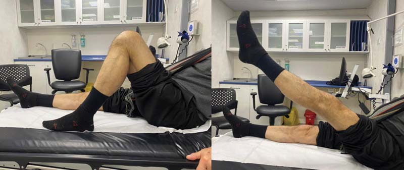
The patient evolved favorably, without complications in the immediate postoperative period. The postoperative control radiograph showed adequate reduction and fixation ([Figure 3]). Orthoses were not used postoperatively. The patient began rehabilitation with a range of motion progressive to tolerance without restriction, and gait with partial weight bearing of the operated limb and use of two canes for one month. Kinesitherapy was started the day after surgery, with a frequency of at least three sessions per week. In the first postoperative control at two weeks of evolution, the patient achieved passive range of motion of 0° to 90° without pain and achieved extended leg elevation without inconvenience ([Figure 4]). The patient evolved asymptomatic, with full range of motion (0° to 135°) at 2 months of evolution. Four months after the surgery, the trophism of the quadriceps muscle was slightly decreased compared to the contralateral muscle, the patient was able to lift his limb with the knee extended against resistance, climb stairs, and perform squats, so he was discharged.


Clinical Case No. 2
A 74-year-old male patient, with no medical history, experienced a fall with a direct hit to the left patella, presenting intense pain, increased volume, and inability to actively extend the knee. The imaging study showed a multifragmentary comminuted articular fracture of the patella with oblique main features, with distraction of the main segments of approximately 15 mm ([Figure 5]).

Definitive surgery was performed two weeks after the accident. With the patient in the supine position, an anterior incision of approximately 10 cm was made in the midline of the left knee. Dissection by planes until the identification of the comminuted fracture of the patella was performed, followed by resection of non-synthesizable lateral osteochondral fragments. The medial osteochondral fragment was solidified with two 3-mm cannulated headless compression screws (HCSs). Adequate reduction was verified under fluoroscopy and palpation. Three vertical wires were passed through transosseous tunnels to the proximal fragment of the patella. They were then passed through the distal pole. Deep to the patellar tendon, a premolded 2.4/2.7-mm rim locking compression plate (LCP Compact Foot, DePuy Synthes, Raynham, MA, US) was installed in the distal pole of the patella. Wire loops were attached, and the medial and lateral wires were passed through the holes at the ends of the plate. The central wire passed through. Finally, these were knotted ([Figure 6]). Adequate reduction and stability of the tissue in the joint range of 0° to 30° was verified. The retinacula were repaired with Vicryl 1.0.

The patient evolved favorably, without complications in the immediate postoperative period. The postoperative control radiograph showed adequate reduction and fixation ([Figure 7]). The patient began rehabilitation with a range of motion progressive to tolerance and gait with partial weight bearing of the operated limb and two canes since the first postoperative day, maintaining at least three sessions of kinesiotherapy per week. In control at three postoperative months, the patient achieved an active range of motion 0° to 120° without pain ([Figure 8]). The knee CT scan showed signs of advanced consolidation. In the fourth month after surgery, the patient recovered quadriceps strength, managing to go up and down stairs, perform squats, and raise the limb with the knee extended against resistance, for which reason he was discharged.


Discussion
Fractures of the distal pole of the patella directly compromise the knee extensor apparatus and are rarely stable fractures, so their conservative treatment is rare and only reserved for a small proportion of stable and non-displaced fractures.[12]
The surgical treatment of these fractures is mainly based on two possible alternatives. One option is to perform a distal partial patellectomy together with reattachment of the patellar tendon using high-strength sutures and transosseous tunnels. For some time, this alternative was the treatment of choice.[12] [13] [14] However, partial patellectomy involves altered patellar length, decreased quadriceps strength, joint stiffness, and pain in the anterior knee.[13] [15] The tissue formed between the bone and the tendon requires a longer time to heal compared to bone-on-bone tissue, so a longer period of immobilization is often required.[10] [16] [17]
On the other hand, it is feasible to seek to achieve an osteosynthesis that preserves the patellar fragments. There are several fixation techniques described in the literature that seek to preserve the inferior pole of the patella.[9] [13] [18] [19] [20] In the more recent literature,[21] the use of vertical wires and basket plate fixation have been shown to be the most reliable fixation methods.
Wire fixation techniques include the use of vertical wire alone, combined with cerclage,[10] or augmented with Krackow sutures,[6] as exemplified in the first case herein reported. The technique using independent vertical wire loops has been shown[6] [10] [22] [23] to achieve satisfactory results without the concerns of requiring prolonged immobilization, implant rupture, infection, or altered patellar tendon length.
Oh et al.[6] performed reduction and osteosynthesis of comminuted distal pole fractures using augmented vertical wire loops with reinsertion of the patellar tendon using Krackow sutures and transosseous tunnels. In their series of 11 patients, after an average of 13 months of follow-up (range: 10 to 23 months), they did not observe patients with nonunion, reduction loss, or osteosynthesis failure. On average, consolidation was achieved 10 weeks after surgery (range: 8 to 12 weeks). The average range of motion was of 129° (range: 120° to 140°). The authors[6] highlight the usefulness of this technique thanks to its easy execution, stability, and good results.
In 2018, Cho et al.[24] published their technique, in which they perform the fixation of comminuted fractures of the distal pole of the patella using a premolded rim plate augmented with vertical wire sutures, as exemplified in the second case herein reported. In their work, the authors[24] included 13 patients treated with 2.0-mm LCPs (6 patients) or 1.5-mm LCPs (7 patients) augmented with 3 to 5 vertical wire loops. Among the results, an average follow-up of 13.5 months (range: 12 to 23 months) stands out, as well as consolidation in all cases, which presented on average 10 weeks after surgery (range: 8 to 12 weeks). The average range of motion was o 127° (range: 120° to 130°). There were no patients with osteosynthesis failure or loss of reduction. No patient reported symptoms of symptomatic osteosynthesis at the end of the follow-up. The authors[24] concluded that this technique provides safe and effective fixation to treat displaced and comminuted fractures of the distal pole of the patella.
As aforementioned, the osteosynthesis of these fractures can also be performed only with plates. The basket plate was introduced in the clinical practice in 1988. It has a shape that adapts to the geometry of the patellar apex by means of six hooks, and it is fixed with two parallel cancellous screws and two screws placed obliquely. Biomechanically, the basket plate can withstand a maximum traction force by the patellar tendon of over 400 N.[12] [25] Fixation of this plate enables early, unrestricted mobility. Despite this, among its disadvantages are its large size and its location, which requires direct contact with the patellar tendon.[26] Last but not least, this plate is not available in many countries, and most surgeons are inexperienced with its use. Matejčić et al.[26] published their 25-year experience using these plates, in which the follow-up of 98 patients is reported, with excellent and good results in 80 and 18 patients respectively.
Recently, He et al.[12] reported the reduction and osteosynthesis of these fractures using a 2.4-mm rim locked plate prepared and contoured in such a way that it perfectly adapts to the arch of the distal pole of the patella. After achieving an adequate reduction and position of the plate, it is fixed by means of locked screws that join it to the proximal fragment. Basically, this technique differs from the technique reported by Cho et al.[24] (exemplified by the second clinical case herein reported) because, although both groups used a rim locked plate, the technique used by He et al.[12] fixes the plate directly with screws, and does not use vertical wire loops. Although they have reported optimal consolidation, functionality, and range of motion at the end of the follow-up, the same authors[12] warn that the limitations of this technique include the small number of patients included (four cases) and the lack of a biomechanical study to test it.
Conclusion
We herein presented two cases of comminuted fractures of the distal pole of the patella managed with reduction and osteosynthesis of the fragments, thus avoiding partial patellectomy and its possible complications. In both cases, vertical wire loops are used. In one case, the fixation was increased with reinsertion of the patellar tendon using transosseous tunnels and Krackow sutures, while, in the second case, the transosseous tunnels were not used, but a rim plate was added with support at the distal pole of the patella. Both patients evolved satisfactorily, recovering their functionality early and with no complications. Both techniques correspond to management alternatives for patients with comminuted fractures of the distal pole of the patella, especially in those who seek early and accelerated rehabilitation. These surgical alternatives are valid options with favorable results according to the reviewed literature; however the current evidence is controversial, and there is no standardized management that proposes one surgical technique over another for this type of patient.
Conflicto de Intereses
Los autores no tienen conflicto de intereses que declarar.
* Study conducted at Hospital del Trabajador ACHS, Santiago, Región Metropolitana, Providencia.
-
Referencias
- 1 Larsen P, Court-Brown CM, Vedel JO, Vistrup S, Elsoe R. Incidence and Epidemiology of Patellar Fractures. Orthopedics 2016; 39 (06) e1154-e1158
- 2 Boström A. Fracture of the patella. A study of 422 patellar fractures. Acta Orthop Scand Suppl 1972; 143: 1-80
- 3 Melvin JS, Mehta S. Patellar fractures in adults. J Am Acad Orthop Surg 2011; 19 (04) 198-207
- 4 Meinberg EG, Agel J, Roberts CS, Karam MD, Kellam JF. Fracture and Dislocation Classification Compendium-2018. J Orthop Trauma 2018; 32 (Suppl. 01) S1-S170
- 5 Matejcić A, Puljiz Z, Elabjer E, Bekavac-Bešlin M, Ledinsky M. Multifragment fracture of the patellar apex: basket plate osteosynthesis compared with partial patellectomy. Arch Orthop Trauma Surg 2008; 128 (04) 403-408
- 6 Oh H-K, Choo S-K, Kim J-W, Lee M. Internal fixation of displaced inferior pole of the patella fractures using vertical wiring augmented with Krachow suturing. Injury 2015; 46 (12) 2512-2515
- 7 Chang S-M, Ji X-L. Open reduction and internal fixation of displaced patella inferior pole fractures with anterior tension band wiring through cannulated screws. J Orthop Trauma 2011; 25 (06) 366-370
- 8 Carpenter JE, Kasman R, Matthews LS. Fractures of the patella. Instr Course Lect 1994; 43: 97-108
- 9 Saltzman CL, Goulet JA, McClellan RT, Schneider LA, Matthews LS. Results of treatment of displaced patellar fractures by partial patellectomy. J Bone Joint Surg Am 1990; 72 (09) 1279-1285
- 10 Yang KH, Byun YS. Separate vertical wiring for the fixation of comminuted fractures of the inferior pole of the patella. J Bone Joint Surg Br 2003; 85 (08) 1155-1160
- 11 Anand A, Kumar M, Kodikal G. Role of suture anchors in management of fractures of inferior pole of patella. Indian J Orthop 2010; 44 (03) 333-335
- 12 He QF, Pan GB, Yu ZF. et al. Novel Rim Plating Technique for Treatment of the Inferior Pole Fracture of the Patella. Orthop Surg 2021; 13 (02) 651-658
- 13 Veselko M, Kastelec M. Inferior Patellar Pole Avulsion Fractures: Osteosynthesis Compared with Pole Resection. JBJS Essent Surg Tech . 2005 ; os-87(1_suppl_1):113-121.
- 14 Kelly MA, Insall JN. Patellectomy. Orthop Clin North Am 1986; 17 (02) 289-295
- 15 Zhang X, Chen X, Jiang J. et al. [Current situation of surgical treatment of inferior polar fracture of patella]. Zhongguo Xiu Fu Chong Jian Wai Ke Za Zhi Zhongguo Xiufu Chongjian Waike Zazhi Chin J Reparative Reconstr Surg 2010; 24 (04) 492-495
- 16 Petermann J, Ishaque B, Ziring E, Gotzen L. External patellotibial transfixation: indications, operative technique and outcome. Knee 2001; 8 (02) 111-121
- 17 Galla M, Lobenhoffer P. [Patella fractures]. Chir Z Alle Geb Oper Medizen . 2005 ; 76(10):987-997; quiz 998-999.
- 18 Henrichsen JL, Wilhem SK, Siljander MP, Kalma JJ, Karadsheh MS. Treatment of Patella Fractures. Orthopedics 2018; 41 (06) e747-e755
- 19 Buezo O, Cuscó X, Seijas R. et al. Patellar Fractures: An Innovative Surgical Technique With Transosseous Suture to Avoid Implant Removal. Surg Innov 2015; 22 (05) 474-478
- 20 Chang C-H, Chuang H-C, Su W-R, Kuan F-C, Hong C-K, Hsu K-L. Fracture of the inferior pole of the patella: tension band wiring versus transosseous reattachment. J Orthop Surg Res 2021; 16 (01) 365-372
- 21 Patel VR, Parks BG, Wang Y, Ebert FR, Jinnah RH. Fixation of patella fractures with braided polyester suture: a biomechanical study. Injury 2000; 31 (01) 1-6
- 22 Song HK, Yoo JH, Byun YS, Yang KH. Separate vertical wiring for the fixation of comminuted fractures of the inferior pole of the patella. Yonsei Med J 2014; 55 (03) 785-791
- 23 Kang NV, Pendegrass C, Marks L, Blunn G. Osseocutaneous integration of an intraosseous transcutaneous amputation prosthesis implant used for reconstruction of a transhumeral amputee: case report. J Hand Surg Am 2010; 35 (07) 1130-1134
- 24 Cho J-W, Kim J, Cho W-T, Gujjar PH, Oh C-W, Oh J-K. Comminuted inferior pole fracture of patella can be successfully treated with rim-plate-augmented separate vertical wiring. Arch Orthop Trauma Surg 2018; 138 (02) 195-202
- 25 Krkovic M, Bombac D, Balazic M. et al. Modified pre-curved patellar basket plate, reconstruction of the proper length and position of the patellar ligament–a biomechanical analysis. Knee 2007; 14 (03) 188-193
- 26 Matejčić A, Ivica M, Jurišić D, Ćuti T, Bakota B, Vidović D. Internal fixation of patellar apex fractures with the basket plate: 25 years of experience. Injury 2015; 46 (Suppl. 06) S87-S90
Address for correspondence
Publication History
Received: 10 August 2021
Accepted: 18 January 2022
Article published online:
20 May 2022
© 2022. Sociedad Chilena de Ortopedia y Traumatologia. This is an open access article published by Thieme under the terms of the Creative Commons Attribution-NonDerivative-NonCommercial License, permitting copying and reproduction so long as the original work is given appropriate credit. Contents may not be used for commecial purposes, or adapted, remixed, transformed or built upon. (https://creativecommons.org/licenses/by-nc-nd/4.0/)
Thieme Revinter Publicações Ltda.
Rua do Matoso 170, Rio de Janeiro, RJ, CEP 20270-135, Brazil
-
Referencias
- 1 Larsen P, Court-Brown CM, Vedel JO, Vistrup S, Elsoe R. Incidence and Epidemiology of Patellar Fractures. Orthopedics 2016; 39 (06) e1154-e1158
- 2 Boström A. Fracture of the patella. A study of 422 patellar fractures. Acta Orthop Scand Suppl 1972; 143: 1-80
- 3 Melvin JS, Mehta S. Patellar fractures in adults. J Am Acad Orthop Surg 2011; 19 (04) 198-207
- 4 Meinberg EG, Agel J, Roberts CS, Karam MD, Kellam JF. Fracture and Dislocation Classification Compendium-2018. J Orthop Trauma 2018; 32 (Suppl. 01) S1-S170
- 5 Matejcić A, Puljiz Z, Elabjer E, Bekavac-Bešlin M, Ledinsky M. Multifragment fracture of the patellar apex: basket plate osteosynthesis compared with partial patellectomy. Arch Orthop Trauma Surg 2008; 128 (04) 403-408
- 6 Oh H-K, Choo S-K, Kim J-W, Lee M. Internal fixation of displaced inferior pole of the patella fractures using vertical wiring augmented with Krachow suturing. Injury 2015; 46 (12) 2512-2515
- 7 Chang S-M, Ji X-L. Open reduction and internal fixation of displaced patella inferior pole fractures with anterior tension band wiring through cannulated screws. J Orthop Trauma 2011; 25 (06) 366-370
- 8 Carpenter JE, Kasman R, Matthews LS. Fractures of the patella. Instr Course Lect 1994; 43: 97-108
- 9 Saltzman CL, Goulet JA, McClellan RT, Schneider LA, Matthews LS. Results of treatment of displaced patellar fractures by partial patellectomy. J Bone Joint Surg Am 1990; 72 (09) 1279-1285
- 10 Yang KH, Byun YS. Separate vertical wiring for the fixation of comminuted fractures of the inferior pole of the patella. J Bone Joint Surg Br 2003; 85 (08) 1155-1160
- 11 Anand A, Kumar M, Kodikal G. Role of suture anchors in management of fractures of inferior pole of patella. Indian J Orthop 2010; 44 (03) 333-335
- 12 He QF, Pan GB, Yu ZF. et al. Novel Rim Plating Technique for Treatment of the Inferior Pole Fracture of the Patella. Orthop Surg 2021; 13 (02) 651-658
- 13 Veselko M, Kastelec M. Inferior Patellar Pole Avulsion Fractures: Osteosynthesis Compared with Pole Resection. JBJS Essent Surg Tech . 2005 ; os-87(1_suppl_1):113-121.
- 14 Kelly MA, Insall JN. Patellectomy. Orthop Clin North Am 1986; 17 (02) 289-295
- 15 Zhang X, Chen X, Jiang J. et al. [Current situation of surgical treatment of inferior polar fracture of patella]. Zhongguo Xiu Fu Chong Jian Wai Ke Za Zhi Zhongguo Xiufu Chongjian Waike Zazhi Chin J Reparative Reconstr Surg 2010; 24 (04) 492-495
- 16 Petermann J, Ishaque B, Ziring E, Gotzen L. External patellotibial transfixation: indications, operative technique and outcome. Knee 2001; 8 (02) 111-121
- 17 Galla M, Lobenhoffer P. [Patella fractures]. Chir Z Alle Geb Oper Medizen . 2005 ; 76(10):987-997; quiz 998-999.
- 18 Henrichsen JL, Wilhem SK, Siljander MP, Kalma JJ, Karadsheh MS. Treatment of Patella Fractures. Orthopedics 2018; 41 (06) e747-e755
- 19 Buezo O, Cuscó X, Seijas R. et al. Patellar Fractures: An Innovative Surgical Technique With Transosseous Suture to Avoid Implant Removal. Surg Innov 2015; 22 (05) 474-478
- 20 Chang C-H, Chuang H-C, Su W-R, Kuan F-C, Hong C-K, Hsu K-L. Fracture of the inferior pole of the patella: tension band wiring versus transosseous reattachment. J Orthop Surg Res 2021; 16 (01) 365-372
- 21 Patel VR, Parks BG, Wang Y, Ebert FR, Jinnah RH. Fixation of patella fractures with braided polyester suture: a biomechanical study. Injury 2000; 31 (01) 1-6
- 22 Song HK, Yoo JH, Byun YS, Yang KH. Separate vertical wiring for the fixation of comminuted fractures of the inferior pole of the patella. Yonsei Med J 2014; 55 (03) 785-791
- 23 Kang NV, Pendegrass C, Marks L, Blunn G. Osseocutaneous integration of an intraosseous transcutaneous amputation prosthesis implant used for reconstruction of a transhumeral amputee: case report. J Hand Surg Am 2010; 35 (07) 1130-1134
- 24 Cho J-W, Kim J, Cho W-T, Gujjar PH, Oh C-W, Oh J-K. Comminuted inferior pole fracture of patella can be successfully treated with rim-plate-augmented separate vertical wiring. Arch Orthop Trauma Surg 2018; 138 (02) 195-202
- 25 Krkovic M, Bombac D, Balazic M. et al. Modified pre-curved patellar basket plate, reconstruction of the proper length and position of the patellar ligament–a biomechanical analysis. Knee 2007; 14 (03) 188-193
- 26 Matejčić A, Ivica M, Jurišić D, Ćuti T, Bakota B, Vidović D. Internal fixation of patellar apex fractures with the basket plate: 25 years of experience. Injury 2015; 46 (Suppl. 06) S87-S90