Keywords
brain metastases - stereotactic radiosurgery - cumulative intracranial tumor volume
- stereotactic laser ablation
Introduction
Brain metastasis (BM) affects 20 to 40% of cancer patients, translating to approximately
170,000 cases in the U.S. each year.[1] While brain metastases can arise from virtually any cancer, they are most commonly
found in patients afflicted with lung cancer (19.9%), melanoma (6.9%), renal cell
carcinoma (RCC) (6.5%), breast cancer (5.1%), and colorectal cancer (1.8%).[2] Patients with HER2 breast cancer, triple negative breast cancer, melanoma, small
cell lung cancer, and non-squamous/nonsmall cell lung cancer (NSCLC) have the highest
risk of developing brain metastases.[3] The majority of BM patients present with oligometastatic disease, typically defined
as 1 to 3 intracranial lesions.[4] Tumors such as melanoma and RCC have higher propensities for intracranial invasion
relatively early during clinical course while breast and colorectal cancer invade
the central nervous system after systemic metastases have been established.[5]
In this review, we will provide an overview of stereotactic radiosurgery (SRS) as
a treatment for BM. Emphasis will be placed on efficacy, prognostic variables, patient
selection, and complications.
Diagnosis and Prognosis
Any cognitive decline or acute neurological symptom in a patient diagnosed with cancer
merits prompt imaging workup. Approximately 90% of patients with brain metastases
suffer neurocognitive decline prior to diagnosis.[6] For workup, the gold standard diagnostic tool is a thin axial magnetic resonance
imaging (MRI) performed after administration of contrast material. On T1 imaging,
brain metastases are typically solid, contrast-enhancing masses located at the gray-white
junction. Magnetic resonance spectroscopy tends to demonstrate high choline/N-acetylaspartate
and choline/creatinine ratios in the contrast-enhancing regions.[7] BM from melanomas, choriocarcinomas, germ cell tumors, thyroid cancer, and RCC are
more likely to be hemorrhagic.[8]
The prognosis of patients with brain metastases is poor, with a median survival of
4 months after whole brain radiation therapy (WBRT) and 1-year survival of 12%.[9]
[10] However, survivors beyond historical expectations are beginning to emerge as improved
systemic therapy becomes available through targeted agents and immunotherapies.[11]
[12] Clinical variables that prognosticate survival can be divided into three categories:
patient demographics and clinical condition (age, Karnofsky Performance Score [KPS]
greater, systemic disease control), BM characteristics (cumulative intracranial tumor
volume [CITV], number of BM), and the presence of targetable mutations (e.g., BRAF
mutation). The relative importance of these prognostic factors varies as a function
of the specific cancer type[13] and cancer-specific prognostic scales have been developed for BM patients. These
scales aid in clinical decision making in terms of palliative versus curative intent.[14]
Treatment Options
Because most systemic therapies poorly penetrate the blood–brain barrier, their application
as therapies for BM require participation in pertinent clinical trials or the discretion
of the treating oncologist. It is important to note that some BMs do respond to systemic
therapy, particularly for smaller sized lesions.[15]
Surgical resection is considered for patients with oligometastatic disease with symptomatic
mass effect. Resection or biopsy is also warranted in cases of diagnostic uncertainty.[16] Additionally, randomized controlled trials demonstrate improved survival in patients
with solitary BM who underwent surgical resection followed by radiation therapy relative
to those treated with radiation therapy alone.[17]
[18] Importantly, surgical resection should be followed with radiation of the resection
cavity, since approximately 50% of resected tumors recur locally without such treatment.[19]
Radiation therapy remains a mainstay option for the treatment of BM and can be applied
to the entire brain, as WBRT or only to the BM (SRS). In WBRT, radiation is delivered
in small fractions on a daily basis. It is highly effective in achieving local control
of tumor growth.[20] Most studies report local control rate of over 80%.[21]
[22]
[23]
[24] Because the entire cerebrum is radiated, WBRT “sterilizes” regions of the brain
that are not affected by the macroscopic tumor. Since these regions may harbor micrometastatic
foci that were invisible to the original MRI, WBRT minimizes the likelihood of new
BM distant to the original tumor site (termed distant metastasis). This control of
distant metastasis comes at the cost of injury to the cerebrum and neurocognitive
decline following treatment. In two independent clinical trials, oligometastatic BM
patients with WBRT exhibited worsened verbal memory capacity relative to those treated
with SRS.[4]
[25] In general, current clinical practice employs WBRT for: prophylactic cranial irradiation
for small cell lung cancers, treatment of miliary BM, or treatment with palliative
intent.[8]
[26]
SRS involves technology platforms that converge multiple, nonparallel beams to deliver
a single, high radiation dose to a targeted region.[27]
[28] The radiation delivered through SRS is highly conformal to the lesion, with a rapid
dose fall-off at the edge of the treatment volume.[5] With the exception of highly radiation resistant tumors, such as melanomas and sarcomas,
SRS is highly efficacious as a means of controlling BM growth.[29]
[30] Since SRS spares cerebrum unaffected with BM, there is a decreased likelihood of
posttreatment neurocognitive decline relative to WBRT.[4]
[25]
[31] Moreover, because higher doses can typically be delivered through SRS, local control
is improved relative to WBRT.[29]
[30]
[32] However, repeat radiosurgery as treatment for distant recurrence is often required.[30] As such, continued imaging surveillance is required for patients who undergo SRS.
While WBRT and SRS differ in the control of local and distant BM, most studies indicate
comparable survival after either treatment.[4]
[29] These observations are largely consistent with studies demonstrating uncontrolled
systemic disease as the main cause of cancer death.[33]
[34] In this context, while there is an extended literature describing the effects of
combining SRS with WBRT,[4]
[20]
[29]
[35] this practice is not routinely applied in the current clinical practice.
Platforms for SRS
The concept of SRS was first introduced by Leksell in 1951 with the use of several
proton beams and later on the gamma beams.[36] Since this initial landmark development, multiple technology platforms have been
developed to facilitate SRS. These technology platforms bear distinct commercial names,
including Gammaknife,[37] Cyberknife,[38] Edge,[39] Hyperarc,[40] ZAP,[41] and proton beam radiosurgery.[42] While the mechanisms of radiation delivery differ between these platforms, the available
literature suggests comparable clinical efficacy.[43]
Considerations for SRS Treatment
Considerations for SRS Treatment
Dose, Fraction, and Anatomic Considerations
Based on a landmark study by the Radiation Therapy Oncology Group (RTOG), the maximum
tolerated SRS doses for tumors less than 20, 21 to 30, and 31 to 40 mm were 24, 18,
and 15 Gy, respectively.[44] More current clinical applications utilize doses below the thresholds defined by
this study. Dose deescalation when treating lesions in proximity of radiation sensitive
structures, such as the optic nerve and the brainstem are routinely performed.[45]
Historically, SRS requires headframe placement and is typically delivered in a single
treatment. With improvement in methods for immobilization as well as time required
for delivery, frameless SRS is now possible. For larger lesions, patients can undergo
hypofractionated SRS, defined as up to five treatments of conformation radiation delivery.[46] An alternative approach for SRS of larger lesions involves staged SRS, where SRSs
are separated by short time intervals or sequentially delivered to different regions
of the lesion.[47] Efficacy of these treatment variations for larger lesions are largely comparable
to single fraction radiosurgery for smaller lesions. Dose equivalent radiation delivered
through fractionation has been shown to decrease posttreatment morbidity (Lau et al[48]).
Prognostic Scales
Survival prognostication serves as a key foundation for tailoring therapy to BM patients.
Several prognostic scales have been developed for SRS-treated BM patients ([Table 1]). The earlier prognostic scales, including the recursive partitioning analysis (RPA),
modified RPA, Score Index for Radiosurgery, Basic Score for Brain Metastasis, and
Graded Prognosis Analysis (GPA),[48]
[49]
[50]
[51]
[52]
[53] treated BM as a single entity, irrespective of the original cancer diagnosis. These
studies highlight the prognostic importance of patient demographics and clinical condition
as well as BM characteristics. In recent studies, there is increased appreciation
that BM derived from cancers of distinct histology exhibit differing clinical courses[54] and that prognostic scales need to be tailored to distinct tumor types. The disease-specific
GPA was developed in this context ([Table 2]). With the emergence of therapies targeting oncogenic mutations, such as BRAF and
EGFR, modern prognostic scales now incorporate tumor mutation status as a prognostic
factor.[55]
[56]
Table 1
Summary of nontumor-specific prognostic scales
|
Year of publication
|
Number of patients
|
Prognostic variables
|
Resulting parameter
|
Median overall survival (mo)
|
|
Age (y)
|
KPS
|
Primary tumor control
|
Tumor number
|
Non-brain metastases
|
Largest tumor volume (cm3)
|
|
RPA[48]
|
1997
|
1,200
|
< 65
≥ 65
|
≥ 70
< 70
|
Yes
No
|
NA
|
NA
|
NA
|
Class I: Age < 65, KPS ≥ 70, and primary tumor control
Class II: All others
Class III: KPS < 70
|
Class I: 7.1
Class II: 4.2
Class III: 2.3
|
|
Modified-RPA for class II[49]
|
2012
|
3,753
|
NA (All age ≥ 65)
|
0: 90–100
1: 70–80
|
0: Yes
1: No
|
0: Single
1: Multiple
|
0: No
1: Yes
|
NA
|
Class IIa: score of 0–1
Class IIb: score of 2
Class IIc: score of 3–4
|
Class IIa: 19.7–15.6
Class IIb: 8.4
Class IIc: 5.2–3.5
|
|
Modified-RPA for class III[50]
|
2002
|
916
|
< 65
≥ 65
|
NA (All KPS < 70)
|
Yes
No
|
Single
Multiple
|
NA
|
NA
|
Class IIIa: Age < 65, primary tumor control, single lesion
Class IIIb: All others
Class IIIc: Age ≥ 65, uncontrolled primary tumor, multiple lesions
|
Class IIIa: 3.2
Class IIIb: 1.9
Class IIIc: 1.2
|
|
SIR[51]
|
2000
|
65
|
0: ≥ 60
1: 51–59
2: ≤ 50
|
0: ≤ 50
1: 50–70
2: ≥ 70
|
0: PD
1: PR-SD
2: CR-NED
|
0: ≥ 3
1: 2
3: 1
|
NA
|
0: > 13
1: 5–13
2: < 5
|
Score range of 0–10
|
Score 1–3: 2.9
Score 4–7: 7.0
Score 8–10: 31.38
|
|
BS-BM[52]
|
2004
|
110
|
NA
|
0: 50–70
1: 80–100
|
0: No
1: Yes
|
NA
|
0: Yes
1: No
|
NA
|
Score range of 0–3
|
Score 0: 1.9
Score 1: 3.3
Score 2: 13.1
Score 3: Undefined
|
|
GPA[53]
|
2008
|
1,960
|
0: > 60
0.5: 50–59
1: < 50
|
0: < 70
0.5: 70–80
1: 90–100
|
NA
|
0: > 3
0.5: 2–3
1: 1
|
0: Present
1: None
|
NA
|
Score range of 0–4
|
Score 0–1: 2.6
Score 1.5–2.5: 3.8
Score 3: 6.9
Score 3.5–4: 11.0
|
Abbreviations: BS-BM, Basic Score for Brain Metastasis; CR, complete clinical remission;
GPA, Graded Prognosis Analysis; KPS, Karnofsky Performance Score; NA, not applicable;
NED, no evidence of disease; PD, progressive disease; PR, partial remission; RPA,
recursive partitioning analysis, SD, stable disease; SIR, Score Index for Radiosurgery.
Table 2
Summary of tumor-specific prognostic scales
|
Year of publication
|
Number of patients
|
Prognostic variables
|
Resulting parameter
|
Median overall survival (mo)
|
|
Age (y)
|
KPS
|
Tumor number
|
Non-brain metastases
|
Gene status
|
|
ds-GPA (lung - NSCLC and SCLC)[a]
[13]
|
2008
|
4,529
|
0: > 60
0.5: 50–59
1: < 50
|
0: < 70
0.5: 70–80
1: 90–100
|
0: > 3
0.5: 2–3
1: 1
|
0: Present
1: Absent
|
NA
|
Score range of 0–4
|
Score 0–1: 2.79–3.02
Score 1.5–2.5: 5.3–6.5
Score 3: 9.6–11.3
Score 3.5–4: 14.8–17.1
|
|
ds-GPA (lung)[55]
|
2017
|
2,186
|
0: ≥ 70
0.5: < 70
|
0: < 70
0.5: 70–80
1: 90–100
|
0: >4
0.5: 1–4
|
0: Present
1: Absent
|
0: EGFR neg/unk and ALK neg/unk
1: EGFR pos or ALK pos
|
Score range of 0–4
|
AdenoCa
Score 0–1.0: 6.9 Score 1.5–2.0: 13.7
Score 2.5–3.0: 26.5
Score 3.5–4.0: 46.8
|
Non-AdenoCa
Score 0–1.0: 5.3
Score 1.5–2.0: 9.8
Score 2.5–3.0: 12.8.
Score 3.5–4: Unavailable
|
|
ds-GPA (melanoma and RCC)[13]
|
2008
|
4,529
|
NA
|
0: < 70
1: 70–80
2: 90–100
|
0: > 5
1: 2–3
2: 1
|
NA
|
NA
|
Score range of 0–4
|
Score 0–1: 3.3–3.4
Score 1.5–2.5: 4.7–7.3
Score 3: 8.8–11.3
Score 3.5–4: 13.2–14.8
|
|
ds-GPA (melanoma)[56]
|
2017
|
823
|
0: ≥ 70
0.5: < 70
|
0: < 70
0.5: 70–80
1: 90–100
|
0: > 4
0.5: 2–4
1: 1
|
0: Present
1: Absent
|
0: BRAF neg/unk
1: BRAF pos
|
Score range of 0–4
|
Score 0–1.0: 4.9
Score 1.5–2.0: 8.3
Score 2.5–3.0: 15.8
Score 3.5–4.0: 34.1
|
|
ds-GPA (GI and breast)[13]
|
2008
|
4,529
|
NA
|
0: < 70
1: 70
2: 80
3: 90
4: 100
|
NA
|
NA
|
NA
|
Score range of 0–4
|
Score 0–1: 3.1–6.1
Score 1.5–2.5: 4.4–9.4
Score 3: 6.9–16.9
Score 3.5–4: 13.5–18.7
|
Abbreviations: AdenoCa, adenocarcinoma; ALK, anaplastic lymphoma kinase; ds-GPA, diagnosis-specific
Graded Prognostic Assessment; EGFR, endothelial growth factor; GI, gastrointestinal;
KPS, Karnofsky Performance Score; NA, not applicable; neg/unk, negative/unknown; NSCLC,
nonsmall cell lung cancer; pos, positive; RCC, renal cell carcinoma; SCLC, small cell
lung cancer.
a Same scale as original GPA.
Published studies suggest other variables that warrant consideration as prognostic
factors. In one study of lung cancer BM, tumors that were more spherical in morphology
were associated with better local control after SRS.[57] In another study, a pretreatment biological measure, the neutrophil-to-lymphocyte
ratio, appeared to be predictive of local failure after SRS and poor survival.[58] Finally, radiomic features of NSCLC BM have also been associated with prognosis.
A study analyzing 576 NSCLC brain metastases in 161 patients treated with SRS identified
select radiomic features of BM on MRI that were associated with clinical survival.[59] Future prognostic scales should consider incorporation of these variables.
Number of Brain Metastases
Number of Brain Metastases
The Congress of Neurological Surgeons (CNS) 2019 Guidelines for the use of SRS for
the treatment of metastases in adults[60] provide level 3 recommendations for SRS as treatment of patients presenting with
2 to 4 BM.
This recommendation should be considered in the context of the study by Yamamoto et
al, who conducted a prospective study of 1,194 SRS-treated BM patients. While the
overall survival for patients with one BM (13.9 months) was improved relative to those
with 2 to 10 BM (10.8 months), there were no significant differences in overall survival
between those with 2 to 4 BM and those with 5 to 10 BM.[61] In the largest retrospective study to date, Ali et al[63] analyzed 5,750 patients treated with SRS for BM and recapitulated the findings reported
by Yamamoto et al.[62] Other studies have reported similar findings.[30]
[63]
[64] Moreover, in patients with more than 4 lesions, there is level 3 evidence for the
use of SRS to improve overall survival when the cumulative volume is less than 7 mL.[60] These studies support consideration for SRS in the treatment of more than 4 BM in
select circumstances. The findings that lesion size,[63]
[64] patient KPS,[30] and tumor histology[13]
[63] influence local control following SRS treatment of multiple BM bear relevance to
this decision.
Cumulative Intracranial Tumor Volume
Cumulative Intracranial Tumor Volume
CITV is defined as the sum of the volume of all BM detected at the time of diagnosis.
It is an important prognostic factor for patients afflicted with SRS. In general,
maximal radiation dose that can be safely delivered during SRS is inversely proportional
to the volume of the BM. As such, dose deescalation is often required in the treatment
of BM with larger CITV.[44] Moreover, BM with larger CITV is more likely to be associated with mass effect,
which portends to poor prognostication. Finally, larger CITV may reflect an aggressive
biology, which necessarily impacts survival prognostication.[65]
[66]
The CNS 2019 Guidelines for the use of SRS for the treatment metastases in adults[60] provide level 3 recommendations for the use of SRS in the treatment of BM with cumulative
volume of less than 7 mL. However, it is important to keep in mind that the prognostic
threshold for CITV differs depending on the cancer histology. For instance, the prognostic
CITV threshold for colorectal BM is 10 mL while the prognostic CITV threshold is 4 mL
for lung cancer, melanoma, and RCC. In contrast, CITV does not influence survival
in breast cancer who undergo SRS treatment for BM.[67] In this context, the 7-mL guideline is more of an “average” of the prognostic threshold
for the various cancer types. It is important to bear this observation in mind when
considering the CNS guidelines.
Additionally, it is essential to recognize that because of the heterogeneity in the
volume of BMs, the number of BM does not always correlate with the CITV. In multiple
studies, CITV and number of BM constitute independent prognostic factors.[45]
[65]
[68]
[69] Thoughtful consideration of these variables in the context of cancer histology,
patient condition, and molecular profile of the BM is warranted.
Complications
Recurrent Brain Metastasis
Depending on tumor histology and CITV, up to 30% of SRS-treated BM recur. Although
repeated SRS treatments of these lesions are feasible, they are associated with increased
risk for radiation necrosis (RN) and treatment failure.[70] As such, stereotactic laser ablation (SLA) represents an alternative therapeutic
option. SLA involves the insertion of a fiber-optic probe into the lesion, followed
by laser activation to trigger thermocoagulation ([Fig. 1]). As a stand-alone treatment for BM that recur after SRS, local control is largely
influenced by the percent of tumor ablation.[71] Tumor recurrence is more likely when the ablation is incomplete.[72] In completed ablated BM, reported local control is consistently above 80%.[74] However, incomplete ablation can be safely combined with repeat, hypofractionated
SRS to improve local control.[72] Importantly, SLA has been shown to improve quality of life for patients suffering
from BM that recurred after SRS.[73]
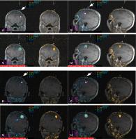 Fig. 1 Thermometry of stereotactic laser ablation. Panels (A) (preablation magnetic resonance imaging [MRI]) and (C) (thermometry) represent coronal images; (B) (preablation MRI) and (D) (thermometry) demonstrate sagittal images. Panels (E) (preablation MRI) and (G) (thermometry) represent coronal images of the same patient in a second ablation;
panels (F) (preablation MRI) and (H) (thermometry) demonstrate sagittal images. Temperature gradient demonstrated as
heat maps. Orange pixels indicate regions of irreversible thermal damage. Arrows indicate insertion site. Asterisks indicate probe tip location.
Fig. 1 Thermometry of stereotactic laser ablation. Panels (A) (preablation magnetic resonance imaging [MRI]) and (C) (thermometry) represent coronal images; (B) (preablation MRI) and (D) (thermometry) demonstrate sagittal images. Panels (E) (preablation MRI) and (G) (thermometry) represent coronal images of the same patient in a second ablation;
panels (F) (preablation MRI) and (H) (thermometry) demonstrate sagittal images. Temperature gradient demonstrated as
heat maps. Orange pixels indicate regions of irreversible thermal damage. Arrows indicate insertion site. Asterisks indicate probe tip location.
Distant Recurrence
Although SRS provides good local tumor control, lesions distant to the original treated
site can develop. In patients who suffer distant BM after initial SRS, the duration
between the two treatments is approximately 6 months.[45]
[70]
[74] Additionally, approximately 20% of patients require more than one additional SRS
as treatment of distant BM. The actuarial freedom from progression of the retreated
tumors at 52 weeks is 92.4%[74] and local control rate at 6 months after salvage SRS is 90.7%.[70] Importantly, patients who undergo 1, 2, 3, 4 or greater repeat SRS exhibit comparable
survival, suggesting efficacy of treatment. Notably, patients who received repeated
SRS were more likely to be younger, have control of systemic disease, have metastases
with smaller cumulative total volume, and suffer from melanoma, indicating a bias
in patient selection for consideration of repeat SRS.[45] Repeat SRS of distant lesions improves quality of life for treated patients in terms
of improving neurologic function as well as discontinuation of corticosteroids.[75]
Multiple repeat SRS treatments, however, are associated with a risk for neurologic
morbidity, including radionecrosis, nonspecific fluid-attenuated inversion recovery
(FLAIR) signal abnormalities, cyst formation seizures, and hemorrhage.[63]
[76]
[77]
[78]
[79]
Leptomeningeal Disease
Leptomeningeal disease involves the seeding of BM cells along the pia mater, arachnoid
mater, and the cerebrospinal fluid (CSF)-filled subarachnoid space. It is a dreaded
complication of cancer, with an extremely poor prognosis of survival that ranged 8
weeks to 6 months.[3] There has been no definitive conclusion as to whether SRS increases the risk of
leptomeningeal disease. However, a recent systematic review suggests patients treated
with SRS are at increased risk for developing leptomeningeal disease when the primary
tumor histology was breast cancer.[80] Several studies have compared outcomes of SRS alone with resection surgery + SRS
and identified an increased risk of leptomeningeal disease in the former.[81]
[82]
[83] The underlying premise is that surgical manipulation disperses cancer cells into
the CSF space. Incidence of leptomeningeal disease after surgical resection with or
without postresection SRS ranges 10 to 20%.[58]
[81] The type of leptomeningeal disease that occurs after surgery, however, appears to
differ from that which develops without surgery. On MRI, leptomeningeal disease after
surgical resection tends to appear nodular in contrast to the classic “sugar-coating.”
Moreover, the nodular leptomeningeal disease is associated with improved overall survival.[58]
[84]
There is an increasing number of studies suggesting that the risk of leptomeningeal
after surgery can be mitigated by neoadjuvant SRS prior to surgical resection[85]
[86]
[87]
[88] ([Table 3]). Reported incidence of leptomeningeal disease for patients treated with neoadjuvant
SRS followed by surgery ranged from 5 to 10%. Further investigation into this novel
paradigm is warranted.
Table 3
Neoadjuvant versus adjuvant SRS
|
Year of publication
|
Type of study and cohort size
|
Groups
|
BM characteristics
|
Outcomes
|
|
Local recurrence
|
Overall survival
|
Radiation necrosis incidence
|
Leptomeningeal disease
|
|
Prabhu et al[17]
|
2017
|
Retrospective, 213 (223 BM)
|
1. SRS alone (n = 157)
2. Gross total resection + SRS (n = 63 neoadjuvant, n = 94 adjuvant)
|
Large BM (≥ 4 cm3)
|
1-year recurrence rate
1. 36.7%
2. 20.5%
|
2 year OS rate
1. 19.8%
2. 38.9%
|
1 year rate
1. 12.3%
2. Neoadjuvant: 5%, Adjuvant: 22.6%
|
1 year rate
1. 1.9%
2. 5.8%[a]
|
|
Mahajan et al[18]
|
2017
|
Randomized controlled, 132
|
1. Resection alone (n = 68)
2. Gross total resection + adjuvant SRS (n = 64)
|
Resection of 1–3 BM, resection cavity diameter of ≤ 4 cm
|
1-year recurrence-free rate
1. 43%
2. 72%
|
Median OS time
1. 18 months
2. 17 months[a]
|
NA
|
1 year rate
1. 16%
2. 28%[a]
|
|
Johnson et al[83]
|
2016
|
Retrospective, 330
|
1. SRS alone (n = 218)
2. Gross total resection + adjuvant SRS (n = 112)
|
1–4 BM
|
NA
|
Median OS time
1. 10.6 months
2. 12.9 months[a]
|
NA
|
1 year rate
1. 5.2%2. 16.9%
|
|
Prabhu et al[87]
|
2018
|
Retrospective, 117 (125 BM)
|
Neoadjuvant SRS + gross total resection
|
70% of patients with 1 BM
|
1-year recurrence rate: 19.9%
2-year recurrence rate: 25.1%
|
Median OS time: 17.2 months
OS 2-year rate: 36.7%
|
1 year rate
5.1%
2 year rate
8.1%
|
1 and 2 year rate: 4.3%
|
|
Patel et al[88]
|
2016
|
Retrospective, 180
|
1. Neoadjuvant SRS + resection (n = 66)
2. Resection + adjuvant SRS (n = 114)
|
90% of patients had 1–3 BM
|
1-year recurrence rate
1. 15.9%
2. 12.6%[a]
|
Median OS time
1. 17.1 months
2. 13.5 months
|
2 year rate
1. 4.9%
2. 16.4%
|
2 year rate
1. 3.2%
2. 16.6%
|
|
Patel et al[89]
|
2018
|
Retrospective, 12
|
Neoadjuvant SRS + surgical resection
|
Median tumor diameter: 3.66 cm
|
6-month tumor control rate: 81.8%
1-year tumor control rate: 49.1%
|
OS rate at 6 months: 83.3%
OS rate at 1 year: 74.1%
|
No evidence of RN
|
1 year rate
16% (n = 2)
|
Abbreviations: BM, brain metastasis; NA, not applicable; OS, overall survival; RN,
radiation necrosis; SRS, stereotactic radiosurgery.
a Nonsignificant differences between groups.
Radiation Necrosis and Other Complication from SRS
RN is a poorly defined term that refers to MR changes occurring at or in proximity
to a BM treated by SRS or WBRT. These findings can include new regions of contrast
enhancement, increased FLAIR abnormality, or a combination of both.[31]
[89] Histological features of RN include coagulative and liquefactive necrosis of the
white matter, thickened hyalinization of vessels, as well as a variable density of
reactive cells and inflammatory cells.[90] While some tumor cells may be present, the threshold for determining active tumor
versus RN is poorly defined and vary widely between pathologists.
The true incidence of RN following SRS for BM is difficult to estimate given the lack
of a standardized definition.[89]
[90] Reported incidence ranges from 5 to 30%.[9]
[31]
[63]
[70] Most RN occur within 2 years of SRS,[44] though delayed RN decades after SRS has been reported.[91] Most RN are not associated with neurologic deterioration, though up to 54% of patients
with RN may be symptomatic.[92] Risk factors for SRS-induced RN include radiation dose,[44]
[63] repeat SRS,[70] and tumor mutations.[92]
Patients with asymptomatic RN are monitored with surveillance imaging.[90] Symptomatic RN are typically managed with corticosteroid treatment.[89] For patients whose RN symptoms are refractory to corticosteroid treatment, bevacizumab
therapy,[93] hyperbaric oxygen,[94]
[95] surgical resection,[15]
[96] or SLA are considered.[97] Of these treatments, laser ablation shows tremendous promise in terms of efficacy.
In independent studies involving retrospective and prospective design, SLA has significant
steroid-sparing effects on symptomatic RN.[73]
[97]
[98]
[99] More than 85% of the RN treated with SLA resolves on subsequent MRI[75]
[100]
[101]
[102]
[103]
[104]
[105]([Table 4]).
Table 4
Treatment of radiation necrosis with SLA—summary of studies reporting outcomes for
radiation necrosis due to SRS-treated brain tumors
|
Study
|
Year of publication
|
Cohort
|
Lesion description
|
Local control
|
Overall survival
|
RN response
|
Resolution of symptoms
|
Steroid use
|
Complications
|
|
Ahluwalia et al[75]
|
2018
|
42
|
19 patients with RN (confirmed with biopsy), median lesion volume 6.4 cm3
|
PFS of 74% at 26 weeks
|
72% at 26 weeks (82.1% for RN subgroup)
|
100% (4/4) of RN treated lesions (total SLA ablation) showed complete response
|
NA
|
37% reduced or stopped steroid use at 12 weeks
|
NA
|
|
Rao et al[99]
|
2014
|
15
|
Intracranial recurrent enhancing lesions (BM or RN). Average lesion size was 3.7 cm3
|
PFS of 75.8% at 24 weeks
|
57% at 37 weeks
|
NA
|
5/7 symptomatic patients had resolution/decrease of symptoms
|
NA
|
1 asymptomatic hemorrhage and 1 neurological deficit
|
|
Hong et al[101]
|
2019
|
75
|
33 patients with RN, 18 treated with SLA, mean volume of 6.29 cm3, all lesions were 100% ablated
|
87.8% local PFS at 1 year
|
69% at 1 year
|
NA
|
87% improved in symptoms
|
34.8% weaned off steroids
|
NA
|
|
Rammo et al[102]
|
2018
|
10
|
All patients with biopsy confirmed RN, 86% lesion volume ablated
|
NA
|
64.8% at 1 year
|
69% with RN volume decrease at 6 months
|
NA
|
7/10 weaned off steroids
|
4 patients with neurological deficit
|
|
Smith et al [103]
|
2016
|
25
|
All patients with biopsy confirmed grade 3 and 4 RN
|
PFS time: 9.1 months
|
OS time: 39.2 months
|
5/15 with mean 26.2% decrease in size at 6 months
|
NA
|
3/7 weaned off steroids
|
NA
|
|
Chaunzwa et al[105]
|
2018
|
30
|
Intracranial recurrent enhancing lesions (BM or RN). 19 lesions were biopsy-confirmed
RN
|
NA
|
26.1% at 1 year
|
77% decrease in FLAIR volume at 6 months
|
32% with resolution of symptoms
|
63% weaned off steroids
|
NA
|
Abbreviations: BM, brain metastasis; FLAIR, fluid attenuated inversion recovery; NA,
not available; OS, overall survival; PFS, progression-free survival; RN, radiation
necrosis; SLA, stereotactic laser ablation; SRS, stereotactic radiosurgery.
Conclusion
BM is a frequent sequela of systemic cancer. For patients with oligometastatic BM,
SRS is preferred to WBRT given the deleterious effects of the latter on neurocognition.
Optimizing clinical decisions in SRS-treated BM patients requires reliable prognostication
through synthesis of information pertaining to the condition of the patient, the characteristics
of the tumor, and the availability of efficacious systemic therapy. Thoughtful consideration
in terms of the number of metastasis and CITV is warranted when considering SRS. Local
recurrence, distant recurrence, leptomeningeal disease, and RN present challenges
in SRS-treated BM patients. Emerging literature suggests new technology platforms,
including stereotactic laser thermal ablation,[72] show promise in navigating these challenges.


