

Subscribe to RSS
DOI: 10.1055/s-0043-1771516
A Retrospective Observational Study of Facial Dog Bite Injuries and Its Management in a Tertiary Care Center
Authors


Abstract
Background: Facial dog bite injuries result in significant emotional, psychological, and physical trauma to the victims involved and should be considered a significant health issue. The purpose of this study is to share our experience in the management and to add to the existing medical literature regarding the epidemiological patterns of facial dog bite injuries.
Materials and Methods: This is a single-center retrospective observational study conducted at Dr. RML Hospital, New Delhi, from January 2021 to January 2022. A total of 105 patients were included. The wounds were managed according to the recommendations made by the national rabies control program and surgical intervention was performed in the form of primary suturing or flap cover.
Results: Children of age group 0 to 10 years are most commonly affected. Pet dogs are the cause in 61% of cases and 57.1% of bites were provoked. Midface is most commonly affected and modified Lackmann's class 3A and 3B are the most common wounds.
Conclusion: In view of raising incidence of dog bites with pet dogs, the general public needs to be educated regarding the practices to prevent these injuries. Postexposure prophylaxis should be given to all affected individuals irrespective of the vaccination status of the dog. Immediate surgical intervention gives better results.
Introduction
The number of animal bites reported under the integrated disease surveillance project has increased from 42 lakhs in 2012 to 74 lakhs in 2020 according to data published by the National Centre of Disease Control, Government of India. These bites include bites from animals such as dog, cat, and monkeys that require rabies postexposure prophylaxis. Among these animal bites, dog bites are the leading cause and in 2015 the estimated number of human rabies cases is 20,847. Almost all the cases of rabies in India are due to stray dogs, which act as a reservoir for the disease. Even though the stray dog vaccination program has been started in many parts of India, a significant number of stray dogs are unvaccinated against rabies.
The majority of facial dog bites affect the pediatric population.[1] The predisposing factors are the small stature of the children, the disproportionate size of the head relative to the body, their willingness to bring their face close to the animal, and limited motor skills to defend themselves and escape. The force delivered by an adult dog jaw can be as high as 450 lbs/sq inch.[2] Such a high quantum of force delivered by the sharp teeth of these mammals can result in three main types of soft tissue wounds—punctures, lacerations, and avulsions with or without actual tissue defect. The mechanism of dog bite injury involves an initial bite with a puncture-type wound with pulling force by a dog, and the reflex mechanism of the victim to get away from the animal acts synergistically, leading to the tearing of the soft tissue calling this “hole and tear” effect.[3] Some degree of crush injury also occurs due to the dynamics of the bite.
Facial dog bite injuries result in significant emotional, psychological, and physical trauma to the victims involved[4] and should be considered a significant health issue. The purpose of this study is to share our experience in the management of facial dog bite wounds and to add to the existing medical literature regarding the epidemiological patterns of facial dog bite injuries.
Materials and Methods
This is a single-center retrospective observational study conducted at Dr. RML Hospital, New Delhi, from January 2021 to January 2022. A total of 105 patients presented to the Department of Plastic Surgery with facial dog bite injuries. All the cases were treated as emergency and followed up for 12 months. All facial dog bites presented within 24 hours were included and nonfacial dog bites were excluded from the study.
Relevant patient history was obtained and clinical examination was done and the wounds were classified according to modified Lackmann's classification[3] ([Table 1]). Tetanus toxoid was administered according to the vaccination protocol. All facial dog bites come under category 3 wounds and are managed according to the recommendations made in the national rabies control program published by the National Centre for Disease Control, India, in 2015. Postexposure prophylaxis includes wound management, rabies immunoglobulin, and antirabies vaccine. This is followed for both vaccinated/unvaccinated or provoked/unprovoked bites.
|
Class |
Clinical finding |
Frequency |
Percentage |
|---|---|---|---|
|
I |
Superficial injury without muscle involvement |
16 |
15.2 |
|
IIA |
Deep injury with muscle involvement |
12 |
11.4 |
|
IIB |
Full-thickness injury of cheek/lip with oral mucosal involvement (through-and-through wound) |
12 |
11.4 |
|
IIIA |
Deep injury with tissue defect (complete avulsion) |
25 |
23.8 |
|
IIIB |
Deep avulsion injury exposing nasal or auricular cartilage |
36 |
34.3 |
|
IVA |
Deep injury with severed facial nerve or parotid duct, or both |
3 |
2.9 |
|
IVB |
Deep injury with concomitant bony fracture |
1 |
1.0 |
The dosage for rabies immunoglobulin is equine rabies immunoglobulin 40 IU per kg body weight of the patient and human rabies immunoglobulin 20 IU per kg body weight. The calculated amount of immunoglobulin was infiltrated into and around the wound. Purified chick embryo cell vaccine was given for active immunization according to the updated Thai red cross schedule (2-2-2-0-2). Reconstituted vaccine (0.5 mL) per site (on two sites per visit, one on each deltoid area) was injected intradermally on day 0, 3, 7, and 28.
All the cases were operated by the senior MCh resident doctor or the consultant on duty. The wound management includes thorough irrigation of the wound with normal saline and meticulous debridement and primary suturing, flap cover or skin grafting as per the requirement. The wounds in the territory of the parotid duct were managed with cannulation of the parotid duct and injection of diluted hydrogen peroxide or normal saline and checked for any bubbling and formation of foam to rule out parotid duct injury.[5] All patients were followed up and postoperative complications were managed accordingly.
Results
Out of 105 patients, 37.1% patients belong to the age group 0 to 10 years, 20% patients belong to the age group 11 to 20 years, 20% patients belong to the age group 21 to 30 years, 10.5% patients belong to 31 to 40 years, and 12.4% patients belong to 41 to 50 years. About 67.6% of patients are male and 32.4% of patients are females.
Pet dogs are the cause in 61% of patients and stray dogs in 39% of patients. Indian pariah is the most common cause followed by German Shepard, Pomeranian, Labrador, Rottweiler, Pitbull, and Indian Mastiff with frequencies of 55.2, 16.2, 12.4, 8.6, 3.8, 2.9, and 1%, respectively.
About 57.1% of patients had bite while playing with a dog (provoked). About 18.1% of the patients had bites spontaneously (unprovoked). About 13.3% of the patients had bites while taking selfie videos or photos. About 11.4% of the patients had bites when they are alcohol intoxicated and stepped or fell over the dog. About 53.3% of the patients presented to the hospital within 12 hours from the time of the incident and 46.7% of the patients presented after 12 hours.
The most common sites involved in facial dog bites are cheek, lips, ear, and nose with 32.4, 29.5, 19.0, and 13.3% incidence, respectively ([Fig. 1]). The number of wounds ranged from 1 to 4 with a negative correlation between age and the number of wounds, that is, as the age increases, the number of wounds decreases. Modified Lackmann's classification 3A and 3B is the most common. ([Figs. 2],[3],[4]).


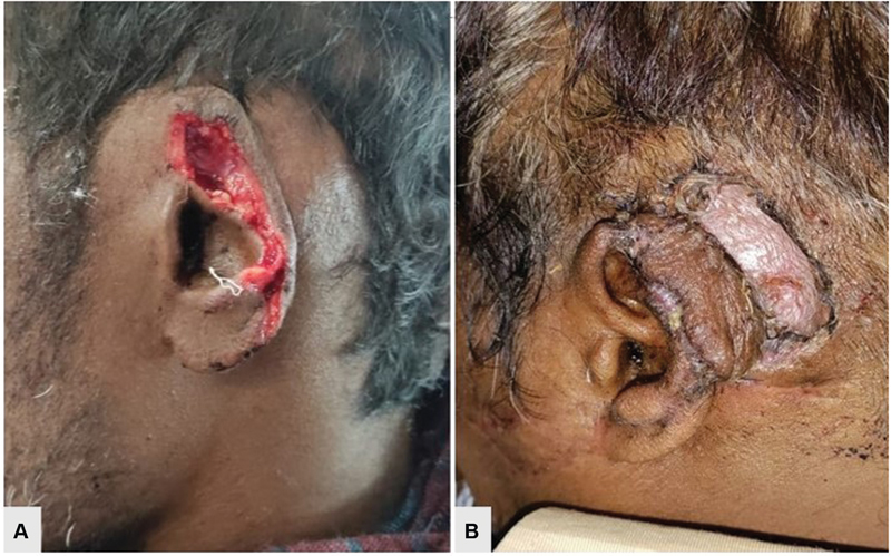

About 85.7% of the patients' wounds were closed primarily with simple suturing, and in 8.6% of the cases flap cover was done, while in 5.7% of cases, skin grafting was done. Out of nine patients who underwent flap cover, three cases were nose defects covered with paramedian forehead flap, bilobed flap, and the banner flap, respectively, while ear defects were managed by postauricular flap in two cases and Antia-Buch helical advancement flap in one case. Lip defects with loss of vermilion and part of the orbicularis oris are managed by mucosal advancement flap. A 3-year-old child who presented with cheek avulsed laceration due dog bite was managed by cheek rotation flap and it is complicated by partial flap necrosis ([Table 2]) that was managed with debridement and full-thickness skin graft in the second stage.
Abbreviation: SSI, surgical site infections.
In 61.0% of the patients, there were no complications. Wound infections and partial flap loss were noted in 21.9% and 1% of cases, respectively. All are superficial surgical site infections (SSI) and hypertrophic scarring was noted in 16.2% of cases
On correlation analysis between the breed of the dog and the age group affected, it is noted that 50% of victims in age group 0 to 10 are affected by Indian pariah (stray dog; [Table 3]). On correlation analysis between the breed of the dog and gender of the victim affected, it is noted that all the bites by Indian pariah (stray dogs) and ferocious dog breeds like German Shepard, Pitbull, and Rottweiler show a clear predominance of males being affected. No correlation between complications and breed is noted in our study. It is noted that complications like SSI, hypertrophic scarring, and flap necrosis are high in cheek injuries.
Discussion
Facial dog bites are a serious public health concern because of infection, scarring, and facial deformity and associated psychosocial effects. Pediatric age group is most commonly affected as described by many studies in the literature.[1] [6] [7] In our study also age group of 0 to 10 years is most commonly affected. Children of this age group will not be able to interpret the warning signs of aggression such as growling, piloerection, and raising the ears and tail.[8] Males are most commonly affected (67.6%) with male to female ratio of 2:1. This could be due to more exposure of males to the outer environment as compared to the women. This finding is similar to previous studies.[9]
Pet dogs are the cause in 61% of the cases as they are cared for close to the face and activities like hugging and kissing the dogs are mostly seen with pet dogs. And this can be contributed to anthropomorphism practice too when treating a dog like a child consists of impeding their physical activity and movement; for example, holding pets on one's lap, carrying them in one's arms or school bags, or transporting them in strollers designed for babies for long periods.[10] These practices can affect the behavior and welfare of companion dogs, by reducing their freedom of movement and consequently their ability to control environmental stimuli. These results are consistent with other studies.[11]
All the bites by stray dogs are by Indian pariahs and in pet dogs German Shepherd is the most common cause followed by Pomeranian, Labrador, and Rottweiler, etc.; other studies also found German Shepherds a common cause for facial bites.[7] [12] Dog breeds like German Shepard, Pitbull, and Rottweilers are ferocious. In a few parts of India, dog breeds like Pitbull, Rottweiler, Tibetan Mastiff, and American Bulldog are banned in view of public safety.
Most of the bites are provoked (57.1%) due to falling over the dog, stepping over the dog's tail, or hugging the dog. In recent times, the increased use of social media and taking selfie videos and photos with the face close to the dog caused facial dog bites in 13.3% of cases. The middle third of the face is frequently involved in our study, reflecting the finding of Palmer and Ress, who called this the central target area.[8] The most common sites involved are the cheek, lips, ear, and nose and these findings are consistent with Berzon.[13] Berzon explained that facial bites are an extension of the face and mouth-biting behavior of dogs with each other.
Considering the robust vascularity of the face, the wound is closed primarily in 85.7% of the case. Flap or graft cover is considered only in cases of gross tissue loss or avulsion injury with devitalized skin. No wounds were left to heal by secondary healing. In one case of cheek laceration that was managed by cheek rotation flap was complicated by flap necrosis, which was debrided and resurfaced with full thickness skin grafting in the second stage. Out of three patients with type 4a injury, one had parotid duct injury that is repaired after stenting and two patients had facial nerve injury medial to the vertical line from lateral canthus; hence, no repair is done considering the dense branching and both cases showed complete recovery in 6 months.
Dog bites are considered untidy wounds as they are contaminated with the oral bacterial flora of the dog, and wound infection is the most common complication following these injuries and the estimated rate of infection is up to 25%.[14] SSI was noted in 21.9% of cases and all are superficial SSI. On detailed analysis, the following predisposing factors for the SSIs are noted that include bite by a stray dog, time of presentation more than 12 hours from the time of the incident, and deep avulsion injury exposing nasal or auricular cartilage (3b). All the cases were managed with local wound dressings and antibiotics cover and no patient developed rabies or life-threatening infection during the treatment or in follow-up.
Conclusion
Facial dog bite injuries are increasing especially with the pet dogs. Pediatric age group is the most commonly affected. Close supervision of child dog interactions, discouraging provocative practices, better reporting of dog bites at various levels, maintaining dog registry of breeds in the community, and dog bite awareness programs involving health care workers will help in preventing these injuries. Postexposure prophylaxis should be given to all affected individuals irrespective of the vaccination status of the dog. Immediate surgical intervention of facial dog bite injuries gives better results.
Conflict of Interest
None declared.
-
References
- 1 Karlson TA. The incidence of facial injuries from dog bites. JAMA 1984; 251 (24) 3265-3267
- 2 Natarajan S, Galinde JS, Asnani U, Sidana S, Ramaswami R. Facial dog bite injury. J Contemp Dent 2012; 2 (02) 34-38
- 3 Williams JV, Magennis P, Graham AJ. Modification of Lackmann's classification of facial bite wounds: an aid to assessment and recording. Br J Oral Maxillofac Surg 2018; 56 (09) 900-901
- 4 Ali SS, Ali SS. Dog bite injuries to the face: a narrative review of the literature. World J Otorhinolaryngol Head Neck Surg 2022; 8 (03) 239-244
- 5 Lawson III GA, Kreymerman P, Nahai F. An unusual complication following rhytidectomy: iatrogenic parotid injury resulting in parotid fistula/sialocele. Aesthet Surg J 2012; 32 (07) 814-821
- 6 Jain P, Jain G. Study of general awareness, attitude, behavior, and practice study on dog bites and its management in the context of prevention of rabies among the victims of dog bite attending the OPD services of CHC Muradnagar. J Family Med Prim Care 2014; 3 (04) 355-358
- 7 Piccart F, Dormaar JT, Coropciuc R, Schoenaers J, Bila M, Politis C. Dog bite injuries in the head and neck region: a 20-year review. Craniomaxillofac Trauma Reconstr 2019; 12 (03) 199-204
- 8 Agrawal A, Kumar P, Singhal R, Singh V, Bhagol A. Animal bite injuries in children: review of literature and case series. Int J Clin Pediatr Dent 2017; 10 (01) 67-72
- 9 Touré G, Angoulangouli G, Méningaud JP. Epidemiology and classification of dog bite injuries to the face: a prospective study of 108 patients. J Plast Reconstr Aesthet Surg 2015; 68 (05) 654-658
- 10 Mota-Rojas D, Mariti C, Zdeinert A. et al. Anthropomorphism and its adverse effects on the distress and welfare of companion animals. Animals (Basel) 2021; 11 (11) 3263
- 11 Schultz RC, McMaster WC. The treatment of dog bite injuries, especially those of the face. Plast Reconstr Surg 1972; 49 (05) 494-500
- 12 Jain R, Prakash G, Midya M, Sharma P. Diverse spectrum of facial dog bite presentation and their management. International Surgery Journal 2018; 5 (09) 3017-3022
- 13 Berzon DR. The animal bite epidemic in Baltimore, Maryland: review and update. Am J Public Health 1978; 68 (06) 593-595
- 14 Rothe K, Tsokos M, Handrick W. Animal and human bite wounds. Dtsch Arztebl Int 2015; 112 (25) 433-443
Address for correspondence
Publication History
Article published online:
04 August 2023
© 2023. Association of Plastic Surgeons of India. This is an open access article published by Thieme under the terms of the Creative Commons Attribution-NonDerivative-NonCommercial License, permitting copying and reproduction so long as the original work is given appropriate credit. Contents may not be used for commercial purposes, or adapted, remixed, transformed or built upon. (https://creativecommons.org/licenses/by-nc-nd/4.0/)
Thieme Medical and Scientific Publishers Pvt. Ltd.
A-12, 2nd Floor, Sector 2, Noida-201301 UP, India
-
References
- 1 Karlson TA. The incidence of facial injuries from dog bites. JAMA 1984; 251 (24) 3265-3267
- 2 Natarajan S, Galinde JS, Asnani U, Sidana S, Ramaswami R. Facial dog bite injury. J Contemp Dent 2012; 2 (02) 34-38
- 3 Williams JV, Magennis P, Graham AJ. Modification of Lackmann's classification of facial bite wounds: an aid to assessment and recording. Br J Oral Maxillofac Surg 2018; 56 (09) 900-901
- 4 Ali SS, Ali SS. Dog bite injuries to the face: a narrative review of the literature. World J Otorhinolaryngol Head Neck Surg 2022; 8 (03) 239-244
- 5 Lawson III GA, Kreymerman P, Nahai F. An unusual complication following rhytidectomy: iatrogenic parotid injury resulting in parotid fistula/sialocele. Aesthet Surg J 2012; 32 (07) 814-821
- 6 Jain P, Jain G. Study of general awareness, attitude, behavior, and practice study on dog bites and its management in the context of prevention of rabies among the victims of dog bite attending the OPD services of CHC Muradnagar. J Family Med Prim Care 2014; 3 (04) 355-358
- 7 Piccart F, Dormaar JT, Coropciuc R, Schoenaers J, Bila M, Politis C. Dog bite injuries in the head and neck region: a 20-year review. Craniomaxillofac Trauma Reconstr 2019; 12 (03) 199-204
- 8 Agrawal A, Kumar P, Singhal R, Singh V, Bhagol A. Animal bite injuries in children: review of literature and case series. Int J Clin Pediatr Dent 2017; 10 (01) 67-72
- 9 Touré G, Angoulangouli G, Méningaud JP. Epidemiology and classification of dog bite injuries to the face: a prospective study of 108 patients. J Plast Reconstr Aesthet Surg 2015; 68 (05) 654-658
- 10 Mota-Rojas D, Mariti C, Zdeinert A. et al. Anthropomorphism and its adverse effects on the distress and welfare of companion animals. Animals (Basel) 2021; 11 (11) 3263
- 11 Schultz RC, McMaster WC. The treatment of dog bite injuries, especially those of the face. Plast Reconstr Surg 1972; 49 (05) 494-500
- 12 Jain R, Prakash G, Midya M, Sharma P. Diverse spectrum of facial dog bite presentation and their management. International Surgery Journal 2018; 5 (09) 3017-3022
- 13 Berzon DR. The animal bite epidemic in Baltimore, Maryland: review and update. Am J Public Health 1978; 68 (06) 593-595
- 14 Rothe K, Tsokos M, Handrick W. Animal and human bite wounds. Dtsch Arztebl Int 2015; 112 (25) 433-443