

Subscribe to RSS
DOI: 10.1055/s-0045-1806769
Bone Density Improvement with Croton lechleri in Alveolar Preservation Procedures: A Tomographic Evaluation Pilot Study
Authors

Abstract
Objectives
This study aimed to evaluate the effect of Croton lechleri on alveolar preservation, focusing on density, dimensions, epithelial regeneration, and pain scale.
Materials and Methods
This pilot randomized clinical trial included 20 patients aged 35 to 56, divided into two groups: Group A (experimental) and Group B (control). Both underwent atraumatic dental extractions in the anterior maxilla. Group A received C. lechleri in the alveolus post-extraction, while Group B received no treatment. Patients with <25% buccal wall loss and defects <3 mm were randomized using block randomization via Jamovi software. Assignments were sealed in opaque envelopes and revealed after data collection. Cone-beam computed tomography and clinical analyses were conducted over 4 months.
Statistical Analysis
For the statistical analysis of tomographic data, an analysis of variance test for repeated measures was used, followed by a post hoc Tukey test. The clinical data analysis was conducted using a Student's t-test.
Results
Exploratory findings from this pilot study revealed a statistically significant effect on bone density (p = 0.03). Although reduced bone loss was noted with the use of C. lechleri extract, the experimental group demonstrated better outcomes in terms of preserving dimensional changes. Furthermore, patients in the experimental group required fewer analgesics during the first 72 hours (p = 0.01), suggesting a significant effect on the clinical pain management protocol and a potential positive effect of C. lechleri on alveolar preservation. In this study, no adverse effects were observed. Regarding the epithelial regeneration parameters, no differences were noted in the color of the scar tissue at 72 hours (p = 1.00). Croton lechleri appears to accelerate the bone repair process, allowing for the formation of higher quality bone in terms of density.
Conclusion
This study demonstrates that the use of C. lechleri is viable for improving bone density, providing better pain control, and achieving favorable epithelial healing. However, larger studies are necessary to confirm these findings.
Keywords
alveolar preservation technique - bone regeneration - cone-beam computed tomography - image J - bone densityIntroduction
During the post-extraction alveolar healing process, a reduction of up to 50% of the original width is observed, with more pronounced resorption in the buccal region compared with the lingual or palatal regions. Alveolar healing generally occurs within the first 3 months, with radiological evidence of maturation typically seen between 3 to 6 months,[1] This process poses a significant challenge in oral rehabilitation, as the resulting lack of bone structure often renders implant-supported rehabilitation unfeasible.
Various methods and techniques for alveolar preservation are currently under investigation to address this issue. Among them, allografts and xenografts have been employed, showing no significant differences in efficacy and demonstrating effectiveness in mitigating resorption.[2] However, these methods are often impractical in developing countries due to their high cost and limited accessibility.
Guided bone regeneration techniques, which aim to reduce bone loss, preserve blood vessels, promote clot formation, and provide growth factors,[3] have shown promise in alveolar preservation. Nonetheless, these methods can lead to complications in the soft tissues, which significantly influence the success of bone regeneration,[4] In this context, traditional medicine offers a viable alternative, as it is typically associated with fewer adverse effects. For instance, Croton lechleri, known for its various biological activities, has demonstrated potential for increasing cell proliferation without carcinogenic activity or tumor-promoting effects.[5] Recent studies have also revealed its ability to induce programmed cell death in cancer cells,[6] further heightening interest in its application for bone regeneration.
The beneficial properties of C. lechleri, including its ability to promote cell proliferation, collagen synthesis, and the preservation of periodontal ligament cell viability,[7] are critical for the alveolar repair process. Both collagen and epithelial formation play fundamental roles in this context.
During dental alveolus healing, reductions in both volume and density are commonly observed. In this regard, C. lechleri exhibits strong wound-healing potential,[8] as well as cicatrizing and anti-inflammatory properties.[7] [9]
Additionally, tension-free closure, a key factor in enhancing vertical preservation,[10] may also be facilitated by the application of this compound. Its antioxidant properties[11] further underscore its potential in promoting healing and bone formation in post-extraction alveoli. However, its clinical effects and impact on bone density remain insufficiently explored.
This study aims to evaluate the efficacy of C. lechleri extract in enhancing alveolar healing following tooth extraction. Dimensional changes, clinical parameters, pain management, and bone density analysis were assessed to determine its potential to improve bone formation and alveolar repair.
This pilot study was conducted on post-extraction alveoli of patients who underwent extraction using the alveolar preservation technique, where positive results were observed in bone density levels in the group of patients who used C. Lechleri extract. Thus, this study evaluated the healing effect of C. Lechleri on alveolar preservation, in terms of bone structure formed, bone density, pain scale, soft tissue healing, and the number of analgesics used.
Materials and Methods
The project was conducted at the dental clinic of the Hermilio Valdizán National University, located in the Pillco Marca district, Huánuco province. This study was experimental, prospective, controlled, and blinded regarding treatment allocation, clinical data recording, and analysis. It followed the method of a randomized, double-blind clinical trial, organized according to the CONSORT guidelines, which are used for this type of study. Randomization was performed as follows: initially, patients requiring extraction of a tooth from the anterior region of the maxilla were subsequently divided into two groups by random assignment of numbers. Subsequently, the division of the groups was performed, using the Jamovi software, to ensure randomization, this selection was placed in envelopes of the same color, and these were sealed until the end of the data collection. Additionally, the principal investigator was unaware of which group each patient belonged to, as this procedure was handled by another member of the research team to ensure concealed group allocation.
The study was conducted on patients who came to the dental clinic of the Hermilio Valdizán National University with a single-rooted incisor, canine, or premolar requiring dental extraction.
The inclusion criteria were male or female patients aged between 18 and 50 years, without systemic diseases (ASA I); patients with an indication for tooth extraction without pathological processes; patients without periodontal disease at the time of extraction; patients with a moderate to thick gingival phenotype; and patients with less than 3 mm or less than 25% loss of the buccal wall. Exclusion criteria: smokers, pregnant or breastfeeding women; patients with controlled systemic diseases; patients diagnosed with or suspected of having diabetes; patients taking chronic or current medications; patients with a history of severe bruxism; patients with alcohol dependence, chronic drug abuse, or psychological disorders; patients with clinically symptomatic or asymptomatic periapical radiolucency. Patients were selected based on the inclusion and exclusion criteria, resulting in a total of 20 patients, divided into two groups: a C. lechleri group and a control group, with 10 patients in each group.
The sample selection was obtained through nonprobabilistic and intentional sampling, such that from all the patients who attended the dental clinic service, the one that best fit the study and aligned with the primary objective of the research was chosen. Additionally, due to the nature and/or characteristics of the type of analysis conducted, it was only possible to perform the study with a limited number of patients.
To conduct this study, observation was used as a technique, both clinically and through imaging exams, collecting data from variables for subsequent analysis. Additionally, surveys were used to gather data on certain specific variables. The instruments used in this research were a data collection form and a questionnaire developed to gather information regarding the tomographic analysis, as well as clinical and symptomatic aspects, for subsequent processing.
Data Processing
After data collection, the data will be processed using the Excel 2019 program, followed by the SPSS statistical package version 26.0 for Windows.
For tomographic analysis, the analysis of variance test for repeated measures was used, followed by Tukey's post hoc test. In all cases, the normality of the data was tested using the Shapiro–Wilk test. For the clinical data analysis, Student's t-test for independent groups was performed.
Tomographic Analysis
Dimensional Evaluation
To assess the dimensions of the alveoli, all patients underwent tomographic examinations (cone-beam computed tomography [CBCT]), performed immediately postoperatively, and a second evaluation was done 4 months after the operation. All tomographic exams were performed using the same device (Soredex, Scanora 3D), adhering to the principles of ALARA (As Low as Reasonably Achievable) for tomography of specific areas. Alveolar measurements were performed using the software CS 3D Imaging, version 3.10.38, in coronal, axial, and sagittal planes with a 1 mm thickness, and were recorded at two time points: immediately and 4 months later ([Fig. 1]). The measurements analyzed were as follows:
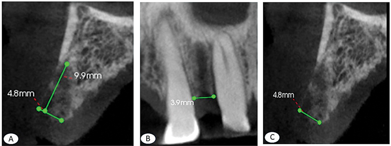
HA: (Height of the socket), the lowest point to the highest crest of the alveolus, perpendicular to EA at 90° ([Fig. 1]).
EA: (Width of the socket), the outermost distance from the buccal cortex to the palatal cortex, in the sagittal plane ([Fig. 1]).
LA: (Length of the socket), the distance from the mesial wall to the distal wall at the highest point of the alveolus, in the frontal section ([Fig. 1]).[12]
All procedures were performed by a single trained dental surgeon (E.U.R.), who was unaware of the group to which each socket belonged, knowing only that the extractions had already been performed.
Density Evaluation:
Relative bone density (RBD) was measured according to the method of Chiapasco et al.[13] This method indicates that the RBD value is obtained by calculating the ratio between the measured density value of the gray area of the bone defect (void or repaired) and the gray value of the surrounding healthy bone area. The software ImageJ (Wayne Rasband, National Institutes of Health; URL: https://imagej.net/ij/download.html; ImageJ packaged with Java 8, 64-bit) was used to perform this analysis. The analysis was conducted in two phases: immediately postoperatively and 4 months postoperatively. The following sequence was followed: (1) capture of tomographic images in sagittal section, 1 mm thick, in TIFF format using the software CS 3D Imaging. (2) Conversion of the grayscale image to color format in ImageJ software. To open the tomography file: File Menu → Open → Select the image. To convert the image: Image Menu → Type → RGB Color, then again Image Menu → Color → Create Composite; once again Image → Color → Split Channels, resulting in three color channels: red, green, and blue. Finally, the blue channel is selected, and the image is saved. (3) Density measurement begins by calibrating the parameters for measurement: Analyze → Set Measurements → Mean gray value, then select Plugins → Macros → Record, immediately select the area to be measured, and finally from the Analyze menu → Measure. The collected data are then processed.
Soft Tissue Healing Analysis
This was conducted through a clinical evaluation of the patients, considering the following criteria: first, the evaluation of the color of the epithelialized healing area, with criteria recognizing the color as pink, erythematous, whitish, or cyanotic at 72 hours, 7 days, and 15 days. Additional criteria included the presence or absence of suppuration, edema, gingival recession, and sensitivity of the adjacent teeth. This evaluation was performed for both groups: the experimental group and the control group. All evaluations were performed by a single, duly trained evaluator who was unaware of which group each patient belonged to.[14]
Pain Intensity Analysis
Pain analysis was conducted using the visual analog scale (VAS), employing an 11-point scale (BS-11)23. It was performed at 6, 12, 24, 48, and 72 hours after the surgical procedure. Patients were instructed to record their pain levels on cards provided to them by marking with a “X.”[15]
Analgesic Use Analysis
To analyze analgesic use, the criteria followed were the recording of the first analgesic taken and the total number of analgesics consumed within the first 72 hours. Patients were instructed to take their first analgesic only when they felt pain, to determine if there was any analgesic effect in the experimental C. lechleri group. Additionally, the number of analgesics consumed within the first 72 hours was recorded to assess whether there was any effect from the element used in the experimental group.[16]
Results
A total of 20 extraction procedures were performed using the alveolar preservation technique on individuals aged between 35 and 56 years, 11 (60%) women and 7 (40%) men. Regarding the type of tooth extracted, the participants in the study who had extractions in the anterior region of the maxilla reported extractions of central incisors, lateral incisors, canines, and first premolars.
Tomographic Analysis
The analysis was conducted through two aspects: dimensional analysis and density analysis.
Dimensional analysis: regarding bone height, thickness, and width, statistical differences were observed between the experimental group and the control group at the initial stages and at 4 months. In all cases, a reduction in values was observed over time. On the other hand, no statistical differences were found for height, thickness, and bone length when comparing the experimental groups ([Table 1]). However, for height and thickness, there was a lower degree of reduction in the values for the experimental group ([Fig. 1]), and only in relation to length was there a slightly greater degree of reduction in values in the experimental group ([Fig. 2]).

Abbreviation: DS, standard deviation.
Note: 95% confidence interval, control, experimental: Croton lechleri.
* Comparison analysis between groups.
Density analysis: regarding density, it was observed that there was an increase in density values over time in both the experimental and control groups; however, it was only significant for the experimental group (p = 0.001). Higher density was also observed for the experimental group, with this difference being statistically significant between the groups ([Table 1] and [Fig. 3]).

Soft Tissue Healing
The aspects analyzed were: first, suppuration at 72 hours and 7 days, with none reported in either group; the second aspect was swelling, also analyzed at 72 hours and 7 days, with no signs of swelling in either group; the third aspect was gingival recession, with no cases occurring in either group; the fourth aspect was sensitivity in the adjacent teeth, with no positive reports on this aspect; the fifth aspect was color, analyzed at 72 hours, 7 days, and 15 days. At 72 hours, a whitish and erythematous color was observed, similar in both groups, with no statistically significant differences. At 7 days, a whitish coloration was more commonly observed in the control group, and continuous pink coloration was observed more in the experimental group, with no statistically significant differences between the groups. At 15 days, the coloration was coral pink for both groups in 100% of the cases. It is worth noting that all recorded colors were considered within the parameters of normal color and normal healing.
Pain Intensity
This aspect was analyzed in two ways: first, using the VAS scale, and second, by measuring the number of analgesics and the timing of the first analgesic consumed by the patients. Regarding the pain scale, no statistically significant differences were observed at any of the evaluated periods (6, 12, 24, and 72 hours) between the experimental and control groups. However, lower values were reported in the experimental group (p = 0.87; [Table 2]). As for the number of analgesics, it was observed that the experimental group used 1 to 2 fewer analgesics in the first 72 hours, and this difference was statistically significant ([Table 2] and [Fig. 1]). The timing of the first analgesic intake was slightly longer in the experimental group; however, this difference was not statistically significant ([Table 2] and [Fig. 1]).
Abbreviation: DS, standard deviation.
Note: 95% confidence interval, control, experimental: Croton lechleri.
Discussion
This pilot study is the first to demonstrate the effect of C. lechleri, resulting in a higher level of bone density, associated with a lower pain scale due to the reduced number of analgesics consumed in the first 72 hours. These results suggest that the use of C. lechleri extract directly in the alveolus was effective in generating alveolar repair with greater bone density, which therefore presents greater resistance.
This pilot study is the first to demonstrate the effect of C. lechleri, resulting in a higher level of bone density, associated with a lower pain scale due to the reduced number of analgesics consumed in the first 72 hours. These results suggest that the use of C. lechleri extract directly in the alveolus was effective in generating alveolar repair with greater bone density, which therefore presents greater resistance.
In this study, it was observed that the average bone height loss was 1.6 mm over 4 months in the anterior region. It is known that the most challenging region is the posterior region of the maxilla, and through a study using CBCT, it was observed that the most common group of defects were horizontal defects, with cases of dehiscence and fenestration occurring at the rates of 2.7 and 3.3%, respectively.[17] In this study, individuals with dentition were analyzed to detect bone defects, which may indicate that in addition to the expected bone loss after tooth extraction, they may also present bone defects, leading to even greater bone loss.
Bone density evaluations in this study reported average values of 0.830 in the experimental group at 4 months and 0.619 immediately after tooth extraction. In comparison, studies using xenogeneic bone grafts as socket fillers reported values of 129 at extraction and 158 at 4 months.[18] These results suggest that C. lechleri may be more effective in enhancing bone density, indicating its potential for improving the bone repair process.
Furthermore, the number of analgesics used in the experimental group was significantly lower than in the control group (p = 0.01). This could be attributed to the anti-inflammatory properties of C. lechleri, as previously reported.[19] Chromatographic studies have shown that this extract contains sesquiterpenes, which inhibit the growth of carcinogenic cells. The experimental group also demonstrated superior bone density (p = 0.03), likely due to the formation of a new bone in the mandible, which typically occurs over 3 months.[20] Changes in the alveoli occur between the first 2 and 12 weeks, with evaluations in this study extending to 4 weeks.[21] These findings suggest that C. lechleri has positive effects, though further studies are needed to confirm these results.
In this study, alveolar preservation demonstrated slightly higher density values with the use of C. lechleri extract up to 60 days. In a previous study on humans involving histological analysis, bone volume was reported at 46% of residual graft after 4 months,[22] which is less than 50%. Additionally, other studies have indicated that bone formation markers are most active within the first 2 months. In the present study, during this early phase, the experimental group exhibited better density values, suggesting that a higher density correlates with a greater bone formation. These observations indicate an apparent improvement in the bone formation process, reflected in a slightly greater bone volume.
This study focused on density parameters and found promising results, especially considering that bone formation within the alveolus was evaluated using only coagulum and C. lechleri. Density values are critical for predicting primary stability and bone quality,[23] which are key parameters in assessing the success of implant treatments. A higher density is generally associated with reduced bone resorption,[24] highlighting the need to explore methods that improve bone quality. Enhancing bone quality will ultimately contribute to the efficacy of implant treatments.
One of the primary limitations of this study is the short duration of the observation period. A long-term study would enable a more comprehensive evaluation of the effects of C. lechleri, as the complete bone repair process is known to take up to 6 months. Additionally, as this was a pilot study, the small sample size represents another limitation, which may have obscured more pronounced positive effects. It is important to note that the participants in this study were Latin individuals in good health, which could influence the generalizability of the findings. Despite the limited sample size, positive outcomes were observed, suggesting that with a larger cohort, more definitive evidence of the effects of C. lechleri extract on the bone repair process could be identified.
Finally, this study holds significant relevance as it demonstrates the evident benefits of C. lechleri extract on bone density and dimensional changes. These findings suggest that this compound could accelerate the bone repair process, leading to higher quality bones in less time. However, it is difficult to generalize these results due to the pilot nature of the study and its small sample size. Nevertheless, this is a highly relevant topic in the field of oral rehabilitation and dental implants.
Conclusion
This pilot study demonstrates that the use of C. lechleri extract is feasible and shows potential effectiveness in enhancing the bone healing process of the post-extraction socket, both by improving bone quality and reducing bone repair time. Larger studies are needed to validate these findings.
Conflict of Interest
None declared.
-
References
- 1 Araújo MG, Silva CO, Misawa M, Sukekava F. Alveolar socket healing: what can we learn?. Periodontol 2000 2015; 68 (01) 122-134
- 2 El-Sioufi I, Oikonomou I, Koletsi D, Bobetsis YA, Madianos PN, Vassilopoulos S. Clinical evaluation of different alveolar ridge preservation techniques after tooth extraction: a randomized clinical trial. Clin Oral Investig 2023; 27 (08) 4471-4480
- 3 Misch CE. Implantes Dentais Contemporaneos. 3rd ed.. St. Louis: Elseiver; 2011
- 4 Lim G, Lin G-H, Monje A, Chan H-L, Wang H-L. Wound healing complications following guided bone regeneration for ridge augmentation: a systematic review and meta-analysis. Int J Oral Maxillofac Implants 2018; 33 (01) 41-50
- 5 Vaisberg AJ, Milla M, Planas MC. et al. Taspine is the cicatrizant principle in Sangre de Grado extracted from Croton lechleri. Planta Med 1989; 55 (02) 140-143
- 6 Tzintzarov A, Boyadzhieva SS, Coelho JAP. et al. Novel insights into the biological activity of Croton lechleri twigs extracts and advancements in their sustainable recovery. Molecules 2024; 29 (17) 4161
- 7 Martins CM, Hamanaka EF, Hoshida TY. et al. Dragon's blood sap (Croton lechleri) as storage medium for avulsed teeth: in vitro study of cell viability. Braz Dent J 2016; 27 (06) 751-756
- 8 Namjoyan F, Kiashi F, Moosavi ZB, Saffari F, Makhmalzadeh BS. Efficacy of Dragon's blood cream on wound healing: a randomized, double-blind, placebo-controlled clinical trial. J Tradit Complement Med 2015; 6 (01) 37-40
- 9 Lopes MI, Saffi J, Echeverrigaray S, Henriques JAP, Salvador M. Mutagenic and antioxidant activities of Croton lechleri sap in biological systems. J Ethnopharmacol 2004; 95 (2–3): 437-445
- 10 Urban IA, Monje A, Nevins M, Nevins ML, Lozada JL, Wang H-L. Surgical management of significant maxillary anterior vertical ridge defects. Int J Periodont Restor Dent 2016; 36 (03) 329-337
- 11 Risco E, Ghia F, Vila R, Iglesias J, Alvarez E, Cañigueral S. Immunomodulatory activity and chemical characterisation of sangre de drago (dragon's blood) from Croton lechleri. Planta Med 2003; 69 (09) 785-794
- 12 Vaquero P, Audisio S, Buey V. Uso del software imageJ para evaluar la reparación radiológica de defectos óseos circulares tratadas con matriz ósea desmineralizada en conejos. Cienc Vet 2022; 24 (02) 1515-1883
- 13 Chiapasco M, Rossi A, Motta JJ, Crescentini M. Spontaneous bone regeneration after enucleation of large mandibular cysts: a radiographic computed analysis of 27 consecutive cases. J Oral Maxillofac Surg 2000; 58 (09) 942-948 , discussion 949
- 14 Seerig LM, Nascimento GG, Peres MA, Horta BL, Demarco FF. Accumulated risk from poverty and tooth loss at 31 years of age: the 1982 live birth cohort in Pelotas, Rio Grande do Sul State, Brazil [in Portuguese]. Cad Saude Publica 2020; 36 (08) e00167619
- 15 Ferreira JPR, Araújo PC, Saliba MTA, Moimaz SAS, Garbin CAS. Perfil de los pacientes atendidos en la Clínica de Implantodoncia de las Facultades Adamantinenses Integradas (FAI). Ciencias la Salud 2015; 13 (02) 233-241
- 16 Pérez Barrero BR, Enríquez Calas D, Perdomo Estrada C, González Rodríguez Wde la C, Noriega Roldán SO. Morbidity in elderly with dental loss. Medisan (Santiago De Cuba) 2020; 24 (03) 381-395
- 17 Ozcan G, Sekerci AE. Classification of alveolar bone destruction patterns on maxillary molars by using cone-beam computed tomography. Niger J Clin Pract 2017; 20 (08) 1010-1019
- 18 Munhoz EA, Ferreira Junior O, Yaedu RYF, Granjeiro JM. Radiographic assessment of impacted mandibular third molar sockets filled with composite xenogenic bone graft. Dentomaxillofac Radiol 2006; 35 (05) 371-375
- 19 Moreno-Quirós CV, García-Escalante V, Sánchez-Medina A. et al. Croton stipulaceus Kunth, a native Mexican medicinal plant with antioxidant and anti-inflammatory activities. Bol Latinoam Caribe Plantas Med Aromat 2024; 23 (04) 523-533
- 20 Thor A, Franke-Stenport V, Johansson CB, Rasmusson L. Early bone formation in human bone grafts treated with platelet-rich plasma: preliminary histomorphometric results. Int J Oral Maxillofac Surg 2007; 36 (12) 1164-1171
- 21 Flügge T, Nelson K, Nack C, Stricker A, Nahles S. 2-Dimensional changes of the soft tissue profile of augmented and non-augmented human extraction sockets: a randomized pilot study. J Clin Periodontol 2015; 42 (04) 390-397
- 22 Minetti E, Palermo A, Berardini M. Comparison of different techniques in post-extractive socket regeneration using autologous tooth graft: histological and clinical outcomes. Eur J Dent 2024; 18 (02) 477-484
- 23 Aksoy U, Eratalay K, Tözüm TF. The possible association among bone density values, resonance frequency measurements, tactile sense, and histomorphometric evaluations of dental implant osteotomy sites: a preliminary study. Implant Dent 2009; 18 (04) 316-325
- 24 Akoğlan M, Tatli U, Kurtoğlu C, Salimov F, Kürkçü M. Effects of different loading protocols on the secondary stability and peri-implant bone density of the single implants in the posterior maxilla. Clin Implant Dent Relat Res 2017; 19 (04) 624-631
Address for correspondence
Publication History
Article published online:
24 April 2025
© 2025. The Author(s). This is an open access article published by Thieme under the terms of the Creative Commons Attribution License, permitting unrestricted use, distribution, and reproduction so long as the original work is properly cited. (https://creativecommons.org/licenses/by/4.0/)
Thieme Medical and Scientific Publishers Pvt. Ltd.
A-12, 2nd Floor, Sector 2, Noida-201301 UP, India
-
References
- 1 Araújo MG, Silva CO, Misawa M, Sukekava F. Alveolar socket healing: what can we learn?. Periodontol 2000 2015; 68 (01) 122-134
- 2 El-Sioufi I, Oikonomou I, Koletsi D, Bobetsis YA, Madianos PN, Vassilopoulos S. Clinical evaluation of different alveolar ridge preservation techniques after tooth extraction: a randomized clinical trial. Clin Oral Investig 2023; 27 (08) 4471-4480
- 3 Misch CE. Implantes Dentais Contemporaneos. 3rd ed.. St. Louis: Elseiver; 2011
- 4 Lim G, Lin G-H, Monje A, Chan H-L, Wang H-L. Wound healing complications following guided bone regeneration for ridge augmentation: a systematic review and meta-analysis. Int J Oral Maxillofac Implants 2018; 33 (01) 41-50
- 5 Vaisberg AJ, Milla M, Planas MC. et al. Taspine is the cicatrizant principle in Sangre de Grado extracted from Croton lechleri. Planta Med 1989; 55 (02) 140-143
- 6 Tzintzarov A, Boyadzhieva SS, Coelho JAP. et al. Novel insights into the biological activity of Croton lechleri twigs extracts and advancements in their sustainable recovery. Molecules 2024; 29 (17) 4161
- 7 Martins CM, Hamanaka EF, Hoshida TY. et al. Dragon's blood sap (Croton lechleri) as storage medium for avulsed teeth: in vitro study of cell viability. Braz Dent J 2016; 27 (06) 751-756
- 8 Namjoyan F, Kiashi F, Moosavi ZB, Saffari F, Makhmalzadeh BS. Efficacy of Dragon's blood cream on wound healing: a randomized, double-blind, placebo-controlled clinical trial. J Tradit Complement Med 2015; 6 (01) 37-40
- 9 Lopes MI, Saffi J, Echeverrigaray S, Henriques JAP, Salvador M. Mutagenic and antioxidant activities of Croton lechleri sap in biological systems. J Ethnopharmacol 2004; 95 (2–3): 437-445
- 10 Urban IA, Monje A, Nevins M, Nevins ML, Lozada JL, Wang H-L. Surgical management of significant maxillary anterior vertical ridge defects. Int J Periodont Restor Dent 2016; 36 (03) 329-337
- 11 Risco E, Ghia F, Vila R, Iglesias J, Alvarez E, Cañigueral S. Immunomodulatory activity and chemical characterisation of sangre de drago (dragon's blood) from Croton lechleri. Planta Med 2003; 69 (09) 785-794
- 12 Vaquero P, Audisio S, Buey V. Uso del software imageJ para evaluar la reparación radiológica de defectos óseos circulares tratadas con matriz ósea desmineralizada en conejos. Cienc Vet 2022; 24 (02) 1515-1883
- 13 Chiapasco M, Rossi A, Motta JJ, Crescentini M. Spontaneous bone regeneration after enucleation of large mandibular cysts: a radiographic computed analysis of 27 consecutive cases. J Oral Maxillofac Surg 2000; 58 (09) 942-948 , discussion 949
- 14 Seerig LM, Nascimento GG, Peres MA, Horta BL, Demarco FF. Accumulated risk from poverty and tooth loss at 31 years of age: the 1982 live birth cohort in Pelotas, Rio Grande do Sul State, Brazil [in Portuguese]. Cad Saude Publica 2020; 36 (08) e00167619
- 15 Ferreira JPR, Araújo PC, Saliba MTA, Moimaz SAS, Garbin CAS. Perfil de los pacientes atendidos en la Clínica de Implantodoncia de las Facultades Adamantinenses Integradas (FAI). Ciencias la Salud 2015; 13 (02) 233-241
- 16 Pérez Barrero BR, Enríquez Calas D, Perdomo Estrada C, González Rodríguez Wde la C, Noriega Roldán SO. Morbidity in elderly with dental loss. Medisan (Santiago De Cuba) 2020; 24 (03) 381-395
- 17 Ozcan G, Sekerci AE. Classification of alveolar bone destruction patterns on maxillary molars by using cone-beam computed tomography. Niger J Clin Pract 2017; 20 (08) 1010-1019
- 18 Munhoz EA, Ferreira Junior O, Yaedu RYF, Granjeiro JM. Radiographic assessment of impacted mandibular third molar sockets filled with composite xenogenic bone graft. Dentomaxillofac Radiol 2006; 35 (05) 371-375
- 19 Moreno-Quirós CV, García-Escalante V, Sánchez-Medina A. et al. Croton stipulaceus Kunth, a native Mexican medicinal plant with antioxidant and anti-inflammatory activities. Bol Latinoam Caribe Plantas Med Aromat 2024; 23 (04) 523-533
- 20 Thor A, Franke-Stenport V, Johansson CB, Rasmusson L. Early bone formation in human bone grafts treated with platelet-rich plasma: preliminary histomorphometric results. Int J Oral Maxillofac Surg 2007; 36 (12) 1164-1171
- 21 Flügge T, Nelson K, Nack C, Stricker A, Nahles S. 2-Dimensional changes of the soft tissue profile of augmented and non-augmented human extraction sockets: a randomized pilot study. J Clin Periodontol 2015; 42 (04) 390-397
- 22 Minetti E, Palermo A, Berardini M. Comparison of different techniques in post-extractive socket regeneration using autologous tooth graft: histological and clinical outcomes. Eur J Dent 2024; 18 (02) 477-484
- 23 Aksoy U, Eratalay K, Tözüm TF. The possible association among bone density values, resonance frequency measurements, tactile sense, and histomorphometric evaluations of dental implant osteotomy sites: a preliminary study. Implant Dent 2009; 18 (04) 316-325
- 24 Akoğlan M, Tatli U, Kurtoğlu C, Salimov F, Kürkçü M. Effects of different loading protocols on the secondary stability and peri-implant bone density of the single implants in the posterior maxilla. Clin Implant Dent Relat Res 2017; 19 (04) 624-631