

RSS-Feed abonnieren
DOI: 10.1055/s-0045-1814414
Risk Factors for Clavicle Refracture after Plate Removal: A Systematic Review and Meta-Analysis
Autor*innen


Abstract
Although uncommon, clavicle refracture following plate removal poses a major clinical concern. Identifying risk factors may help guide decisions on patient selection for plate removal. The study aims to systematically review and meta-analyze the available literature on risk factors associated with clavicle refracture after plate removal. We conducted a search in PubMed, Scopus, and ScienceDirect and included studies that compared patients with and without refracture after clavicle plate removal. Risk factors were pooled using odds ratios (OR) and standardized mean differences (SMDs), with a random or fixed-effects model depending on heterogeneity. The Newcastle-Ottawa Scale was used to appraise the quality of studies. Five retrospective studies were included, comprising a total of 1,135 patients who met the inclusion criteria. We found that female gender (OR: 0.30; 95% CI: 0.18–0.51) was associated with a lower observed refracture incidence, although this finding is likely confounded. In contrast, lower body weight SMD: 0.65; 95% CI: 0.28–1.03), smaller clavicle diameter (SMD: 0.57; 95% CI: 0.15–0.98), and shorter clavicle length (SMD: 0.68; 95% CI: 0.26–1.09) were significantly associated with an increased risk of refracture. This is the first meta-analysis to identify risk factors for clavicle refracture following plate removal. These findings may inform cautious risk–benefit discussions but do not support individualized prognostication at this time. Overall, the certainty is low, and these results should be interpreted as hypotheses to be tested in future studies, rather than as definitive predictors to guide clinical decision-making.
Introduction
Clavicle fractures are a common orthopedic injury, particularly affecting young, active individuals.[1] Open reduction with internal fixation (ORIF) has become the standard treatment due to its superior outcomes compared with conservative management.[2] [3] However, with the increasing use of plate fixation, the decision of whether and when to remove the implant has become a growing clinical concern.
Although elective plate removal is often performed for symptomatic relief or patient preference, it is not without risk. One of the most notable complications following implant removal is clavicle refracture.[4] [5] While the overall incidence of refracture is relatively low, it can result in morbidity, prolonged rehabilitation, and, in many cases, necessitate a second surgical intervention. Emerging evidence suggests that several factors may predispose patients to refracture. However, the strength and consistency of these associations remain unclear.[5] [6]
To date, no meta-analysis has examined risk factors for refracture following plate removal. Therefore, a synthesis of available evidence is essential. We aimed to identify and quantify risk factors associated with refracture after clavicle plate removal, providing a comprehensive understanding of patient and surgical characteristics that influence this outcome. The findings may guide clinicians in optimizing patient selection and postoperative care to minimize the risk of refracture.
Methods
Search Strategy, Protocol Registration, and Reporting Guideline
We used PubMed, Scopus, and ScienceDirect Databases to search for studies on clavicle refracture published from inception to May 2025, using the search terms “clavicle” AND “refracture” within the databases' title and abstract settings. An Additional search strategy included reference checking of the included studies and Google search. This study adhered to the PRISMA (Preferred Reporting Items for Systematic Reviews and Meta-Analysis) guidelines, and we also prospectively registered our protocol in PROSPERO (CRD420251083806).[7]
Eligibility Criteria
We used the Populations, prognostic, and outcome approach and searched for studies that investigated risk factors for clavicle refracture. We included studies in which (P) patients with clavicle fractures underwent ORIF with plate fixation followed by plate removal after union, (P) the study reported various risk factors, including patient-specific characteristics such as demographic, lifestyle, and procedural risk factors, (O) that can lead to the events of clavicle refracture. We included original articles, such as randomized and observational studies, and required that each study include both a refracture group and a non-refracture group.
Study Selection
Two reviewers (M.R.G. and G.F.) independently performed the title/abstract screening and full text review using the Covidence reference manager. Any disagreement was resolved through discussion and consensus by a third reviewer (F.F.).
Data Extraction and Quality Assessment
Two independent reviewers (M.R.G. and F.F.) extracted the data, which included study details (such as author, publication year, and study design), patient characteristics information (including sample size, group distribution, mean age, and gender), study conclusions, and Newcastle-Ottawa Scale (NOS) assessments. Risk factors were selected from the eligible studies based on two criteria: they represented a comprehensive range of potential risk factor categories, including anthropometric, iatrogenic, fracture-related, and lifestyle factors; and they were reported in at least two separate studies. The same reviewer also independently assesses the quality of the studies using the NOS tools, and any discrepancies during data extraction or quality assessment were resolved through discussion and consensus with a third reviewer (G.F.).
Statistical Analysis
To evaluate the relationship between prognostic factors and the occurrence of refracture, odds ratios (ORs) were calculated for dichotomous variables and standardized mean differences (SMD) for continuous variables, each with a 95% confidence interval (CI). Statistical heterogeneity among the studies was assessed using Chi-square tests and the I 2 statistic, with I 2 values greater than 50% indicating substantial heterogeneity. In cases of substantial heterogeneity, a random-effects model was applied; otherwise, a fixed-effect model was used. Due to the limited number of included studies (fewer than 10), assessment of publication bias was not performed. All results were illustrated using forest plots, and statistical analyses were performed using Review Manager version 5.4.
Results
Literature Search
Our search identified 160 records through the databases. Sixty-two duplicates were removed before initial screening, and 98 records were screened by reading the titles and abstracts. Of these, 89 records were excluded due to irrelevance to the topic or insufficient data. The full texts of nine articles were examined, and four articles were excluded due to inappropriate outcomes, population, or study design. Finally, five studies were included in the synthesis ([Fig. 1]).

Study Characteristic and Quality Assessment
All five studies included in this review were retrospective studies published between 2019 and 2025. The total sample consisted of 1,135 patients, with 1,073 patients in the non-refracture group and 62 in the refracture group. All studies evaluated patients who underwent plate removal after clavicle union, with four studies focused exclusively on midshaft clavicle fractures, and one study included both midshaft and lateral fractures. The mean age of participants ranged from 33 to 48 years across studies, and the proportion of female patients varied from 13.4 to 34%. The reported refracture rates varied between 1.99 and 7.2%. Quality appraisal using the NOS rated all studies with scores ranging from 7 to 9 ([Table 1]).
|
Author, year |
Study design |
Clavicle location included |
Non-fracture group |
Refracture group |
Mean age, gender (female%) |
Refracture rate |
Conclusion |
NOs score |
|---|---|---|---|---|---|---|---|---|
|
Kessler et al[4] 2025 |
Retrospective |
Midshaft and lateral |
50 |
10 |
40 y, Female (20%) |
NA |
•Longer working length is protective |
9 |
|
Zhu et al[9] 2023 |
Retrospective |
Midshaft |
329 |
23 |
48 y, Female (30.1%) |
6.5% |
•Postmenopause, severe comminuted fracture, and poor reduction were significant risk factors for refracture. |
8 |
|
Tsai et al[6] 2019 |
Retrospective |
Midshaft |
258 |
20 |
40.1 y, Female (31.7%) |
7.2% |
• In females, BMI <22.73 greatly increased risk. |
8 |
|
Park et al[10] 2021 |
Retrospective |
Midshaft |
197 |
4 |
37.8 y, Female (13.4%) |
1.99% |
• Simple (A) or complex (C) fractures had higher refracture risk after implant removal. |
7 |
|
Han et al[8] 2025 |
Retrospective |
Midshaft |
239 |
5 |
NR = 46.5, R = 33, Female (34%) |
3.6% |
• Low bone density at screw holes measured by Remaining Bone Ratio (RBR) and index ofremaining Clavicle intensity (IRCI) on X-ray are strongly linked to refracture. Suggests gray value analysis of screw holes may help predict refracture risk. |
8 |
Abbreviations: BMI, body mass index; NOS, Newcastle Ottawa scale; NR, non-refracture; R, refracture.
Across the five included studies, several risk factors for refracture after plate removal were identified. Female gender and low body mass index (BMI) with a cut-off of <22.73 were associated with an increased risk. Postmenopausal status, severe comminution of the fracture, and poor initial fracture reduction were also found to be risk factors. Radiographic parameters such as reduced bone density at screw holes measured through grayscale analysis emerged as potential predictive indicators. Regarding implant-related factors, a longer working length appeared to have a protective effect. Additionally, both simple (type A) and complex (type C) fracture patterns had a higher likelihood of refracture following plate removal ([Table 1]).
Patient-Related Risk Factor
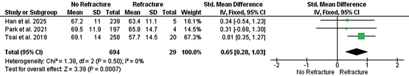
The pooled analysis showed no significant association between age and refracture risk (SMD: 0.25; 95% CI: –0.31–0.81; p = 0.37; I 2 = 59%), In contrast, female gender was significantly associated with reduced risk of refracture occurrence (OR: 0.30; 95% CI: 0.18–0.51; p < 0.0001; I 2 = 0%) ([Fig. 2]), BMI was not found to be a significant risk factor (SMD: 0.29; 95% CI: −0.22–0.80; p = 0.27; I 2 = 51%) No significant associations were found for diabetes (OR: 0.72; 95% CI: 0.20–2.55; p = 0.61; I 2 = not applicable) smoking, or tobacco use (OR: 0.47; 95% CI: 0.20–1.14; p = 0.10; I 2 = 0%), or height (SMD: 0.23; 95% CI: –0.14–0.61; p = 0.23; I 2 = 0%). However, lower body weight was significantly associated with refracture (SMD: 0.65; 95% CI: 0.28–1.03; p = 0.0007; I 2 = 0%) ([Fig. 3]). Menopausal status showed no significant association (OR: 0.52; 95% CI: 0.05–6.02; p = 0.60), but high heterogeneity (I 2 = 84%) limits interpretation. Therefore, female gender was found to have a significantly lower incidence of refracture, and lower body weight was identified as a significant factor for increased risk of clavicle refracture following plate removal ([Table 2]).
Abbreviations: AO/OTA, Arbeitsgemeinschaft für Osteosynthesefragen/Orthopedic Trauma Association; CI, confidence interval.

Biomechanical Risk Factor
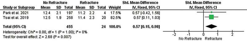
Pooled analyses revealed no statistically significant associations between refracture risk and several fracture classifications, including Robinson type 2A (OR: 2.42; 95% CI: 0.73–8.03; p = 0.15; I 2 = 0%), Robinson type 2B (OR: 0.41; 95% CI: 0.12–1.37; p = 0.15; I 2 = 0%), AO Foundation/Orthopedic Trauma Association (AO/OTA) type A (OR: 0.70; 95% CI: 0.30–1.61; p = 0.40; I 2 = 30%), type B (OR: 1.29; 95% CI: 0.21–7.88; p = 0.78; I 2 = 68%), type C (OR: 0.64; 95% CI: 0.12–3.31; p = 0.60; I 2 = 62%), Robinson type 2B1 (OR: 2.67; 95% CI: 0.28–25.04; p = 0.39; I 2 = 88%), Robinson type 2B2 (OR: 0.24; 95% CI: 0.02–3.03; p = 0.27; I 2 = 91%), and callus formation (OR: 1.48; 95% CI: 0.27–8.08; p = 0.65; I 2 = 0%). Clavicle diameter was significantly smaller in the refracture group (SMD: 0.57; 95% CI: 0.15–0.98; p = 0.007; I 2 = 0%) ([Fig. 4]), indicating it as a risk factor. Additionally, shorter clavicle length was associated with increased risk of refracture (SMD: 0.68; 95% CI: 0.26–1.09; p = 0.001; I 2 = 15%) ([Fig. 5]; [Table 2])

ORIF-Related Risk Factor
The use of interfragmentary or lag screws showed no significant association with clavicle refracture risk (OR: 1.03; 95% CI: 0.58–1.83; p = 0.92; I 2 = 36%), and similarly, the use of wiring was not significantly linked to refracture (OR: 0.74; 95% CI: 0.30–1.84; p = 0.52; I 2 = 17%). Additionally, the time interval between fixation and plate removal was not significantly associated with refracture (SMD: 0.22; 95% CI: −0.15–0.60; p = 0.25; I 2 = 0%).
Discussion
This meta-analysis identified several risk factors for clavicle refracture following plate removal. In pooled analyses, female gender showed a lower observed incidence of refracture after plate removal. This association should not be interpreted as a causal protective effect. It is likely influenced by unmeasured confounding, such as differences in habitual activity levels, return-to-sport profiles, menopausal status, bone health, and case selection for plate removal. Given the observational nature and small sample size, this gender association is best viewed as hypothesis-generating.
Lower body weight was significantly associated with the risk of refracture. Biomechanically, a smaller clavicle diameter and a shorter clavicle length also emerged as risk factors for increased risk of refracture. We found that the types of fracture did not show statistically significant associations, nor did the timing of plate removal. Plate-related variables such as the use of lag screws or wiring showed no meaningful association with refracture incidence.
Our findings somehow differ and at the same time align with those of Tsai et al, who reported that female gender and lower body weight significantly increase the risk of clavicle refracture after plate removal. That study also found low BMI with a cutoff of <22.73, as a risk factor for refracture. However, we found no significant association between BMI and the risk of refracture.[6] Interestingly, four other studies included in this review did not find gender and BMI to be a significant risk factor. At the same time, two of those studies also did not find weight to be a significant risk factor.[4] [8] [9] [10] Although postmenopausal women are theoretically at a higher risk due to estrogen deficiency and reduced bone density, our analysis found no significant associations between menopausal status and refracture, contradicting the findings of Zhu et al. However, our findings are limited by heterogeneity.[9] While body weight showed an association with refracture risk, BMI did not, likely due to BMI's limited ability to accurately reflect body composition. A higher body weight may indicate greater lean muscle and bone mass, whereas lower body weight may contribute to a higher risk of refracture, potentially due to malnutrition, which is commonly associated with being underweight in humans and can lead to osteoporosis. Additionally, factors such as nutrition and menopausal status across populations may further influence the effects of BMI. To better understand these relationships, prospective studies should record actual weight and height, and utilize direct bone density measurements such as Dual-Energy X-ray Absorptiometry (DEXA) scans. Further biomechanical research is needed to confirm these results.[11] [12] Additionally, smoking and diabetes were not associated with refracture in our analysis or in the majority of the included studies. However, due to the small sample size in those studies and in our analysis, which may reduce statistical power, further research is needed to confirm these results.[4] [9] [10]
Our analysis found no significant association between fracture classification (Robinson or AO/OTA) and refracture risk, contradicting Zhu et al and Park et al, who reported associations based on the Robinson and AO/OTA classifications. Similarly, callus formation was not a risk factor in our findings, consistent with two other studies by Tsai et al and Park et al. We also identified a smaller clavicle diameter and shorter clavicle length as significant risk factors for refracture, consistent with the findings in Tsai et al. Larger diameter and longer length may influence bone density, which in turn could be protective for refracture risk.[13] Other studies did not find a significant association between clavicle length or diameter and the risk of refracture.[6] [9] [10]
The use of interfragmentary screws and wiring did not increase the risk of refracture, which is consistent with findings from previous studies suggesting these implant-related factors do not significantly impact outcomes after plate removal. We also found that the time interval between initial plate fixation and hardware removal was not associated with higher refracture risk. These findings are in line with earlier studies that reported no association between the time interval and refracture.[4] [6] [9] [10]
To advance the field, future studies should prioritize prospective multicenter registries with prespecified variable sets, uniform case definitions, standardized follow-up schedules, and preregistered statistical plans that include multivariable adjustment and handling of missing data. Data capture should systematically include key confounders (objective activity exposure, smoking, vitamin D status, comorbidities, bone density via DEXA scans, menopausal status/hormone therapy) and detailed implant and surgical parameters (screw type, interval from fixation to removal, and removal indications). Harmonized outcome reporting, explicit refracture definitions, and minimum follow-up will improve comparability and permit robust sensitivity analyses. Incorporating objective markers of bone healing can guide the timing of implant removal, while data-driven thresholds for BMI, absolute weight, and clavicle dimensions may identify clinically meaningful risk strata.
This meta-analysis has several limitations. First, the number of available studies was small, reducing statistical power and generalizability. Second, we found significant heterogeneity in some of our analyses—such as age, BMI, fracture classification, and menopausal status—which may affect the consistency of pooled results. Third, heterogeneity in surgical technique, rehabilitation protocols, and timing of plate removal across studies could not be fully accounted for. Finally, the evidence remains preliminary, and certainty is low given small, retrospective cohorts, heterogeneity in study designs, inconsistent outcome definitions, and potential confounding.
Conclusion
In conclusion, this meta-analysis identified several significant risk factors for clavicle refracture following plate removal, including female gender, although this factor was associated with lower observed refracture incidence and is likely confounded. In addition, we also found that lower body weight, smaller clavicle diameter, and shorter clavicle length increase the risk of clavicle fracture. Notably, this is the first meta-analysis to explore these risk factors. Given the limited quantity and quality of available evidence, these factors should be considered as possible indicators during shared decision-making about plate removal, but they should not be used as standalone predictors. Until more robust evidence becomes available, discussions with patients should emphasize the overall risk uncertainty and the importance of modifiable factors. Future well-designed, prospective studies are needed to comprehensively evaluate these and other potential risk factors.
Conflict of Interest
None declared.
-
References
- 1 Twomey-Kozak J, Whitlock KG, O'Donnell JA, Klifto CS, Anakwenze O. Epidemiology of sports-related clavicle fractures in the United States: injuries from 2015 to 2019. Orthop J Sports Med 2022; 10 (10) ): 23 259671221126553
- 2 Kassim S, Rai M, Hegde RM, Kishore SG, Shetty S, Balakrishna PN. The study of the functional outcome between surgical and conservatively treated clavicular fractures. Int J Orthopaed Sci 2020; 6: 1205-1209
- 3 Wani MA, Ganaie MA, Islam NUI, Rasool A, Dar NA. Functional results of clavicle fractures in adults treated by open reduction and internal fixation using superior precontoured plate. Int Surg J 2019; 6 (07) 2484-2490
- 4 Kessler F, Kalbas Y, Hambrecht J. et al. Clavicle refractures after hardware removal: are there risk factors? A retrospective cohort study. Eur J Trauma Emerg Surg 2025; 51 (01) 118
- 5 Yin J, Yang G. Refracture after plate removal of midshaft clavicle fracture after bone union during the hospital stay: a case report. Trauma Case Rep 2024; 59: 101100
- 6 Tsai SW, Ma HH, Hsu FW. et al. Risk factors for refracture after plate removal for midshaft clavicle fracture after bone union. J Orthop Surg Res 2019; 14 (01) 457
- 7 Page MJ, McKenzie JE, Bossuyt PM. et al. The PRISMA 2020 statement: an updated guideline for reporting systematic reviews. BMJ 2021; 372: n71
- 8 Han S, Wang Q, Tan F, Li K, Li S. A new method to predict refracture risk after locking compression plate removal of clavicle shaft. BMC Surg 2025; 25 (01) 10
- 9 Zhu Y, Hu J, Zhan T, Zhu K, Zhang C. Refracture after plate removal of midshaft clavicle fractures after bone union-incidence, risk factors, management and outcomes. BMC Musculoskelet Disord 2023; 24 (01) 308
- 10 Park H-Y, Kim S-J, Sur Y-J, Jung J-W, Kong C-G. Refracture after locking compression plate removal in displaced midshaft clavicle fractures after bony union: a retrospective study. Clin Shoulder Elbow 2021; 24 (02) 72-79
- 11 Tanaka S, Kuroda T, Saito M, Shiraki M. Overweight/obesity and underweight are both risk factors for osteoporotic fractures at different sites in Japanese postmenopausal women. Osteoporos Int 2013; 24 (01) 69-76
- 12 Nevitt MC, Cummings SR, Stone KL. et al. Risk factors for a first-incident radiographic vertebral fracture in women > or = 65 years of age: the study of osteoporotic fractures. J Bone Miner Res 2005; 20 (01) 131-140
- 13 Gutiérrez S. CORR Insights®: what regions of the distal clavicle have the greatest bone mineral density and cortical thickness? A cadaveric study. Clin Orthop Relat Res 2019; 477 (12) 2733-2734
Address for correspondence
Publikationsverlauf
Eingereicht: 10. November 2025
Angenommen: 15. November 2025
Artikel online veröffentlicht:
31. Dezember 2025
© 2025. The Author(s). This is an open access article published by Thieme under the terms of the Creative Commons Attribution License, permitting unrestricted use, distribution, and reproduction so long as the original work is properly cited. (https://creativecommons.org/licenses/by/4.0/)
Thieme Medical and Scientific Publishers Pvt. Ltd.
A-12, 2nd Floor, Sector 2, Noida-201301 UP, India
-
References
- 1 Twomey-Kozak J, Whitlock KG, O'Donnell JA, Klifto CS, Anakwenze O. Epidemiology of sports-related clavicle fractures in the United States: injuries from 2015 to 2019. Orthop J Sports Med 2022; 10 (10) ): 23 259671221126553
- 2 Kassim S, Rai M, Hegde RM, Kishore SG, Shetty S, Balakrishna PN. The study of the functional outcome between surgical and conservatively treated clavicular fractures. Int J Orthopaed Sci 2020; 6: 1205-1209
- 3 Wani MA, Ganaie MA, Islam NUI, Rasool A, Dar NA. Functional results of clavicle fractures in adults treated by open reduction and internal fixation using superior precontoured plate. Int Surg J 2019; 6 (07) 2484-2490
- 4 Kessler F, Kalbas Y, Hambrecht J. et al. Clavicle refractures after hardware removal: are there risk factors? A retrospective cohort study. Eur J Trauma Emerg Surg 2025; 51 (01) 118
- 5 Yin J, Yang G. Refracture after plate removal of midshaft clavicle fracture after bone union during the hospital stay: a case report. Trauma Case Rep 2024; 59: 101100
- 6 Tsai SW, Ma HH, Hsu FW. et al. Risk factors for refracture after plate removal for midshaft clavicle fracture after bone union. J Orthop Surg Res 2019; 14 (01) 457
- 7 Page MJ, McKenzie JE, Bossuyt PM. et al. The PRISMA 2020 statement: an updated guideline for reporting systematic reviews. BMJ 2021; 372: n71
- 8 Han S, Wang Q, Tan F, Li K, Li S. A new method to predict refracture risk after locking compression plate removal of clavicle shaft. BMC Surg 2025; 25 (01) 10
- 9 Zhu Y, Hu J, Zhan T, Zhu K, Zhang C. Refracture after plate removal of midshaft clavicle fractures after bone union-incidence, risk factors, management and outcomes. BMC Musculoskelet Disord 2023; 24 (01) 308
- 10 Park H-Y, Kim S-J, Sur Y-J, Jung J-W, Kong C-G. Refracture after locking compression plate removal in displaced midshaft clavicle fractures after bony union: a retrospective study. Clin Shoulder Elbow 2021; 24 (02) 72-79
- 11 Tanaka S, Kuroda T, Saito M, Shiraki M. Overweight/obesity and underweight are both risk factors for osteoporotic fractures at different sites in Japanese postmenopausal women. Osteoporos Int 2013; 24 (01) 69-76
- 12 Nevitt MC, Cummings SR, Stone KL. et al. Risk factors for a first-incident radiographic vertebral fracture in women > or = 65 years of age: the study of osteoporotic fractures. J Bone Miner Res 2005; 20 (01) 131-140
- 13 Gutiérrez S. CORR Insights®: what regions of the distal clavicle have the greatest bone mineral density and cortical thickness? A cadaveric study. Clin Orthop Relat Res 2019; 477 (12) 2733-2734