

Subscribe to RSS
DOI: 10.5999/aps.2015.42.4.489
Giant Extrapleural Solitary Fibrous Tumor of the Thigh
Authors
A solitary fibrous tumor (SFT) is an uncommon tumor that arises from primitive fibroblast-like cells in the connective tissue [[1]]. It characteristically shows a mixture of fibrous tissue, cellular components, and highly vascularized areas consisting of numerous, closely packed small to medium-sized blood vessels [[2] [3]]. They usually affect adults between the fourth and the seventh decades of life (median, 50 years). Histological findings including immunohistochemistry are required for the diagnosis of SFT. Although SFTs are generally benign, well-circumscribed soft tissue tumors, 10%-15% of SFTs will recur and/or metastasize [[4]]. SFTs are usually located in the pleura or other serosal surfaces. Despite the fact that they are seldom located in extrapleural soft tissue, this tumor has been reported in a variety of extraserosal sites. The known extrapleural locations of SFT are as follows: the lumbar extradural space, intrameningeal space, cervical spine, deep soft tissue of the neck, orbital space, pelvic space, retroperitoneal space, vagina, thyroid gland, mammary gland, prostate, nasal mucosa, liver, renal pelvis, and extremities. The orbits and the soft tissues of the extremities are the most commonly reported extrapleural sites [[1]]. SFTs of extremities, particularly in the thighs, are known to have malignant potential [[4]]. To treat benign SFT, resection with an intact tumor capsule is required for full recovery of the patient. Reviewing the literature, we found no confirmed reasons for a wide resection [[4] [5]]. Here, we report a case of an extrapleural SFT that occurred in the inner thigh area.
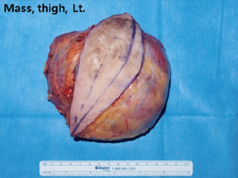
A 76-year-old female presented at our department with a left inner thigh soft tissue mass. Physical examination revealed a huge mass measuring 15 cm×13 cm ([Fig. 1]). Preoperative magnetic resonance imaging (MRI) revealed a large (about 12.4 cm×10 cm), lobulated soft tissue mass with a diffuse hypointense signal on T2-weighted imaging in the subcutaneous fat layer with a bulging contour and peripheral heterogeneous enhancement, which was consistent with a hypervascular tumor ([Fig. 2]). Total body scan revealed no distant metastasis. The mass was located in the subfascial layer of the inner thigh, and surgical treatment was carried out. The whole tumor was totally excised and submitted for histopathological study. The excised specimen was about 15 cm×13 cm in size, encapsulated, and well circumscribed, with a firm, rubbery texture and a tan-brown color ([Fig. 3]). The initial histopathological findings revealed an acellular spindle-cell tumor with nuclear pleomorphism and cellular atypia. They also revealed the proliferation of capillaries surrounded by masses of round or spindle-shaped cells. The cellularity varied considerably in different areas with a predominance of hypercellular areas. However, increased mitotic activity, nuclear pleomorphism, or any foci of coagulative necrosis were not observed in this case. Further, immunohistochemistry was performed for the differential diagnosis. It revealed that CD-34 was positive and S-100 protein was negative ([Fig. 4]). The final histopathological diagnosis was a benign extrapleural SFT. The patient did not receive further radiotherapy or chemotherapy. No recurrence was found 12 months after surgery.


An SFT is a rare neoplasm that derives from mesenchymal cells. The differential diagnosis of an SFT in an extremity includes neoplasms such as fibrosarcoma, fibrous histiocytoma, desmoid tumor, dermatofibrosarcoma protuberans, hemangiopericytoma, neurofibroma, and malignant peripheral nerve sheath tumor [[1]]. Because of the tumor's rarity, it usually takes a long time to reach the diagnosis of SFT. Imaging studies like plain radiography and ultrasound are non-specific and not suitable for the differential diagnosis. On MRI, the diagnosis of SFT is suggested by a well-circumscribed mass with smooth margins, and a focal or diffuse hypointense signal on T2-weighted imaging due to the fibrous content in the tumor. An SFT also demonstrates strong focal or diffuse contrast enhancement due to the highly vascularized areas in the tumor [[1]]. Malignant SFTs are usually demonstrated as hemorrhage, cystic degeneration, and central necrosis on MRI. However, in our case, there was no such evidence suggestive of malignancy. Radiological findings alone cannot definitely determine whether the tumor is benign or malignant [[3]]. To confirm a diagnosis and to differentiate it from other soft tissue tumors, an immunohistochemical analysis is required. SFTs are a well-circumscribed tumors. Histologically, they consist of a proliferation of capillaries surrounded by masses of spindle-shaped cells. SFT cells are separated by thick bands of collagen, demonstrating foci of keloid-like hyalinization. Prominent vascularity showing a hemangiopericytoma-like vascular pattern and thick, hyalinized vessel walls are seen. Immunohistochemically, SFTs are negative for cytokeratin, S-100 protein, desmin, and alpha-smooth muscle actin, while positive for vimentin and CD34 [[2] [3]]. In our case, immunohistochemical staining was positive for CD34 and negative for S-100 protein, which satisfied the diagnostic criteria for SFTs. Patients with a benign SFT are usually treated with complete surgical excision. The prognosis of this tumor is good and the local recurrence rate is very low in the case of benign SFTs. However, other studies have reported that SFTs in the extremities are more likely to be malignant [[4]]. Further, immunohistochemical patterns are used for therapeutic decision making. With mitotic activity, increased cellularity, necrotic areas, and nuclear pleomorphism, there is a possibility of malignant SFT. Thus, if there is evidence suggestive of malignant potential, a further wide resection, a long-term follow-up, and regular MRI are proposed. Otherwise, simple excision with an intact tumor capsule is the optimal treatment of benign SFTs [[4] [5]]. In our case, mitotic activity, nuclear pleomorphism, and central necrosis were not observed. Therefore, simple excision rather than wide excision was sufficient, and there was no evidence of recurrence over the 1 year follow-up period.
In conclusion, although these tumors are an uncommon entity, the possibility of SFTs should be kept in mind during the evaluation of any huge soft tissue mass occurring in the extremities, so that the physician may examine the appropriate differential markers, arrive at an accurate diagnosis, and administer appropriate treatment.
Conflict of Interest
No potential conflict of interest relevant to this article was reported.
-
REFERENCES
- 1 Musyoki FN, Nahal A, Powell TI. Solitary fibrous tumor: an update on the spectrum of extrapleural manifestations. Skeletal Radiol 2012; 41: 5-13
- 2 Hanau CA, Miettinen M. Solitary fibrous tumor: histological and immunohistochemical spectrum of benign and malignant variants presenting at different sites. Hum Pathol 1995; 26: 440-449
- 3 Martorell M, Perez-Valles A, Gozalbo F. et al. Solitary fibrous tumor of the thigh with epithelioid features: a case report. Diagn Pathol 2007; 2: 19
- 4 Anders JO, Aurich M, Lang T. et al. Solitary fibrous tumor in the thigh: review of the literature. J Cancer Res Clin Oncol 2006; 132: 69-75
- 5 Akisue T, Matsumoto K, Kizaki T. et al. Solitary fibrous tumor in the extremity: case report and review of the literature. Clin Orthop Relat Res 2003; (411) 236-244
Correspondence
Publication History
Received: 23 February 2015
Accepted: 04 April 2015
Article published online:
05 May 2022
© 2015. The Korean Society of Plastic and Reconstructive Surgeons. This is an open access article published by Thieme under the terms of the Creative Commons Attribution-NonCommercial License, permitting unrestricted noncommercial use, distribution, and reproduction so long as the original work is given appropriate credit. Contents may not be used for commercial purposes. (https://creativecommons.org/licenses/by-nc/4.0/)
Thieme Medical Publishers, Inc.
333 Seventh Avenue, 18th Floor, New York, NY 10001, USA
-
REFERENCES
- 1 Musyoki FN, Nahal A, Powell TI. Solitary fibrous tumor: an update on the spectrum of extrapleural manifestations. Skeletal Radiol 2012; 41: 5-13
- 2 Hanau CA, Miettinen M. Solitary fibrous tumor: histological and immunohistochemical spectrum of benign and malignant variants presenting at different sites. Hum Pathol 1995; 26: 440-449
- 3 Martorell M, Perez-Valles A, Gozalbo F. et al. Solitary fibrous tumor of the thigh with epithelioid features: a case report. Diagn Pathol 2007; 2: 19
- 4 Anders JO, Aurich M, Lang T. et al. Solitary fibrous tumor in the thigh: review of the literature. J Cancer Res Clin Oncol 2006; 132: 69-75
- 5 Akisue T, Matsumoto K, Kizaki T. et al. Solitary fibrous tumor in the extremity: case report and review of the literature. Clin Orthop Relat Res 2003; (411) 236-244