
RSS-Feed abonnieren
DOI: 10.1055/a-2667-7241
Endoscopic ultrasound-guided pancreatogastrostomy using a novel double lumen dilator


Endoscopic ultrasound-guided pancreatogastrostomy (EUS-PG) can be indicated for patients with pancreatic obstruction after a failed endoscopic retrograde cholangiopancreatography (ERCP) [1] [2] [3]. The technical steps of EUS-PG include pancreatic duct puncture, guidewire deployment, tract dilation, and stent deployment. Notably, compared with the EUS-guided transhepatic approach, the echoendoscope is not stable because its upper angle is not as strong during EUS-PG. During device insertion, such as stent deployment, this instability may lead to inadequate axis and cause performing EUS-PG to be more challenging.
To overcome this problem, a novel double-lumen dilation device (Meissa, Japan Life Line, Tokyo, Japan) was developed ([Fig. 1]). This device has a 2.3-Fr tip and a maximum diameter of 7.4 Fr, with a 2-cm side hole provided from the tip that allows for contrast medium injection, aspiration of the pancreatic juice, and 0.025-in. guidewire insertion. As such, this device can be used to perform the double-guidewire technique without additional device exchange using a 0.018-in. guidewire. Herein, we describe the technical tips for performing EUS-PG using this novel dilator.

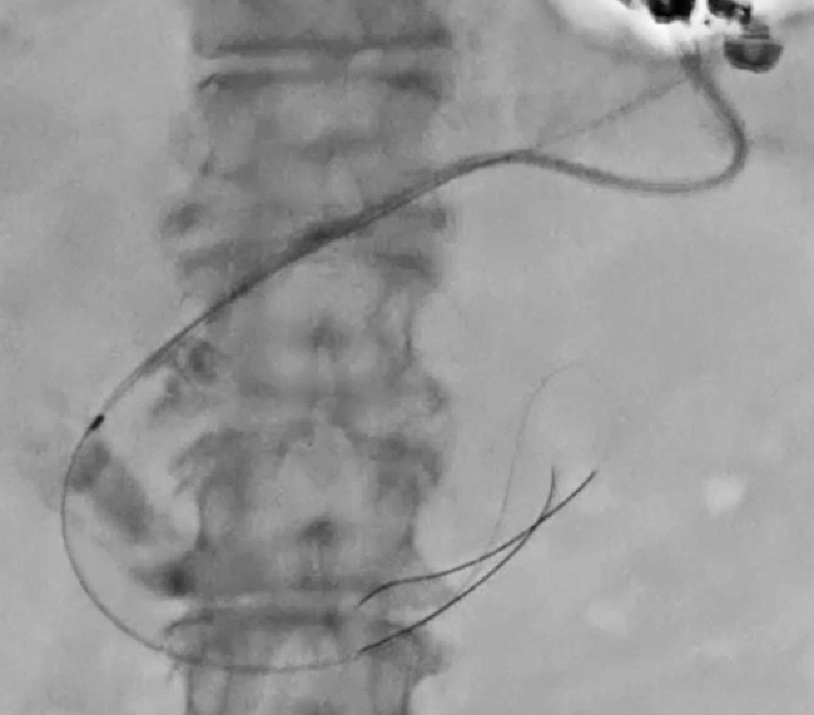
A 78-year-old woman with recurrent pancreatitis due to a pancreatic stone was referred to our hospital after a failed ERCP-guided pancreatic stent deployment at another hospital. Consequently, an EUS-PG was attempted. The pancreatic duct was punctured using a 19-G needle, and contrast medium was injected. A 0.025-in. guidewire was then inserted ([Fig. 2]). Next, the novel dilation device was inserted into the pancreatic tract ([Fig. 3]). A 0.025-in. guidewire was inserted through the side hole of the novel dilator ([Fig. 4]). After tract dilation, a 7.0-Fr stent was easily inserted and successfully deployed from the pancreatic duct to the stomach ([Fig. 5]) without any adverse events ([Video 1]).


In conclusion, this dilation device allows the double-guidewire technique without the need for additional device exchanges and may be useful for EUS-PG.
Endoscopy_UCTN_Code_TTT_1AS_2AD
E-Videos is an open access online section of the journal Endoscopy, reporting on interesting cases and new techniques in gastroenterological endoscopy.
All papers include a high-quality video and are published with a Creative Commons
CC-BY license. Endoscopy E-Videos qualify for HINARI discounts and waivers and eligibility is automatically checked during the submission
process. We grant 100% waivers to articles whose corresponding authors are based in
Group A countries and 50% waivers to those who are based in Group B countries as classified
by Research4Life (see: https://www.research4life.org/access/eligibility/).
This section has its own submission website at https://mc.manuscriptcentral.com/e-videos.
Publikationsverlauf
Artikel online veröffentlicht:
04. September 2025
© 2025. The Author(s). This is an open access article published by Thieme under the terms of the Creative Commons Attribution License, permitting unrestricted use, distribution, and reproduction so long as the original work is properly cited. (https://creativecommons.org/licenses/by/4.0/).
Georg Thieme Verlag KG
Oswald-Hesse-Straße 50, 70469 Stuttgart, Germany
-
References
- 1 Will U, Fueldner F, Buechner T. et al. Endoscopic ultrasonography-guided drainage of the pancreatic duct (EUS-PD)-indications and results with a literature review. J Clin Med 2024; 13: 7709
- 2 Rimbaş M, Larghi A. Endoscopic ultrasonography-guided techniques for accessing and draining the biliary system and the pancreatic duct. Gastrointest Endosc Clin N Am 2017; 27: 681-705
- 3 Teh JL, Teoh AYB. Techniques and outcomes of endoscopic ultrasound guided-pancreatic duct drainage (EUS-PDD). J Clin Med 2023; 12: 1626