

Subscribe to RSS
DOI: 10.1055/a-2751-9622
“Umbrella-shaped pulley traction” assisted endoscopic submucosal dissection of colonic lesions – a modified external traction technique
Authors
Supported by: Scientific Research and Innovation Team of Huai′an First People′s Hospital YCT202305
Supported by: Jiangsu Provincial Medical Key Discipline Cultivation Unit JSDW202233

Colorectal endoscopic submucosal dissection (ESD) is technically demanding because of the difficulty in adequately visualizing the submucosal layer. Therefore, building good traction to provide a clear view for ESD is critically important. Gravity, injection fluid, or a clip-with-line approach has been used to optimize traction during ESD to improve performance [1]. However, the traction direction and ability are limited in most traction methods, resulting in insufficient effects in some cases [2]. Therefore, we adopted a new multi-point and multi-directional external traction method (umbrella-shaped pulley traction) to treat a case of extensive laterally spreading tumors (LSTs) in the rectum ([Video 1]).
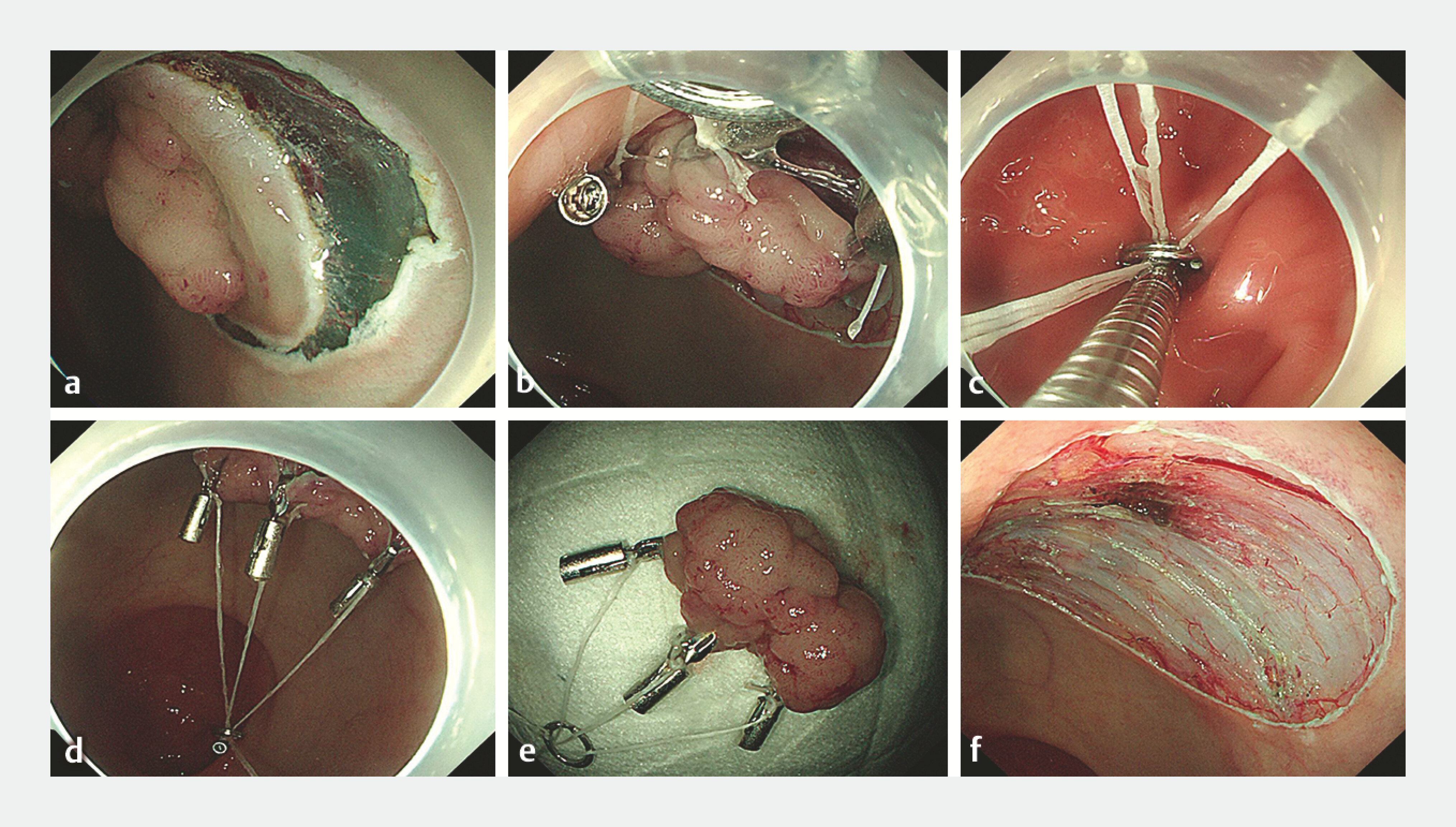
“Umbrella-shaped pulley traction” assisted endoscopic submucosal dissection of an extensive LST in the rectum. LST, laterally spreading tumor.Video 1A 69-year-old woman was found to have a LST (granular type and nodular mixed type) in the rectum. Endoscopic ultrasound indicated that the lesion originated from the mucosal layer and was approximately 2 cm × 3 cm ([Fig. 1]). Therefore, she received ESD for this lesion.

Firstly, a circumferential incision was performed by submucosal injection at the base of the lesion. At the three equal points along the edge of the lesion, a metal clip carrying dental floss was respectively clamped. Subsequently, three pieces of dental floss are placed in a metal ring outside the body. The metal ring was sent into the body with a metal clamp and clamped onto the mucosa on the opposite side of the lesion to form an “umbrella-shaped pulley” structure. The corresponding dental floss can be pulled outside the body as needed to achieve precise traction, allowing the submucosa to be better exposed, providing a clear operational field of view for ESD, and ultimately the lesion is completely removed, ensuring a smooth surgical process ([Fig. 2], [Fig. 3]).

This “umbrella-shaped pulley” traction overcomes the limitations of the previous unidirectional traction, allowing for multi-point and multi-directional traction, which is more convenient to operate. It helps to quickly, completely and safely separate the lesion, especially suitable for larger areas or lesions with low gravity. It is innovative and worth promoting.
Endoscopy_UCTN_Code_TTT_1AQ_2AD_3AD
Contributorsʼ Statement
Rui Xie: Conceptualization. Ru Feng: Writing – original draft. Xiaozhong Yang: Project administration. Honggang Wang: Supervision. Weijie Dai: Writing – review & editing.
Conflict of Interest
The authors declare that they have no conflict of interest.
-
References
- 1 Tziatzios G, Ebigbo A, Glder SK. et al. Methods that Assist Traction during Endoscopic Submucosal Dissection of Superficial Gastrointestinal Cancers: A Systematic Literature Review. Clinical Endoscopy 2020; 53: 286-301
- 2 Nagata M. Advances in traction methods for endoscopic submucosal dissection: What is the best traction method and traction direction?. World J Gastroenterol 2022; 28: 1-22
Correspondence
Publication History
Article published online:
08 January 2026
© 2026. The Author(s). This is an open access article published by Thieme under the terms of the Creative Commons Attribution License, permitting unrestricted use, distribution, and reproduction so long as the original work is properly cited. (https://creativecommons.org/licenses/by/4.0/).
Georg Thieme Verlag KG
Oswald-Hesse-Straße 50, 70469 Stuttgart, Germany
-
References
- 1 Tziatzios G, Ebigbo A, Glder SK. et al. Methods that Assist Traction during Endoscopic Submucosal Dissection of Superficial Gastrointestinal Cancers: A Systematic Literature Review. Clinical Endoscopy 2020; 53: 286-301
- 2 Nagata M. Advances in traction methods for endoscopic submucosal dissection: What is the best traction method and traction direction?. World J Gastroenterol 2022; 28: 1-22